

 PDF
PDF ePub
ePub Citation
 Print
Print


Abstract
Purpose
To Report a case of bilateral central retinal vein occlusion (CRVO) caused by primary antiphospholipid syndrome.
Methods
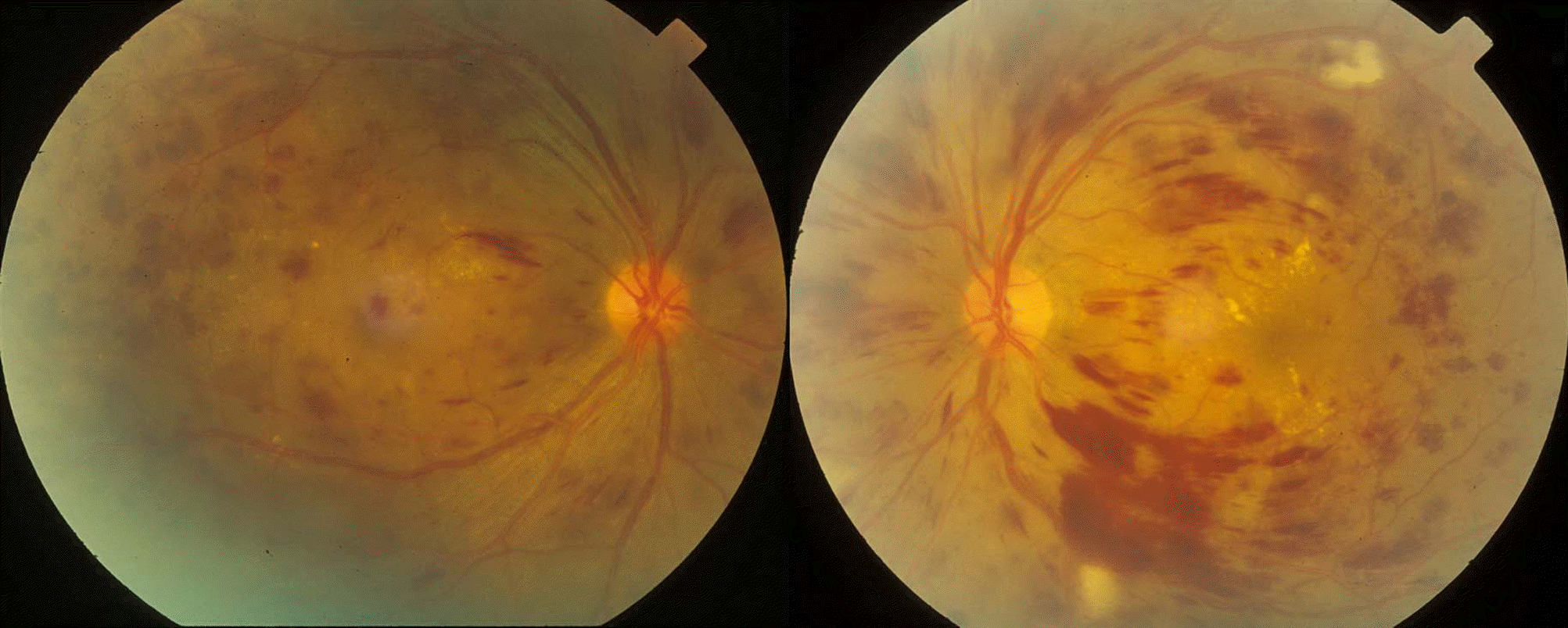
A 38-year-old male is referred to the department of ophthalmology for the bilateral visual loss.
Results
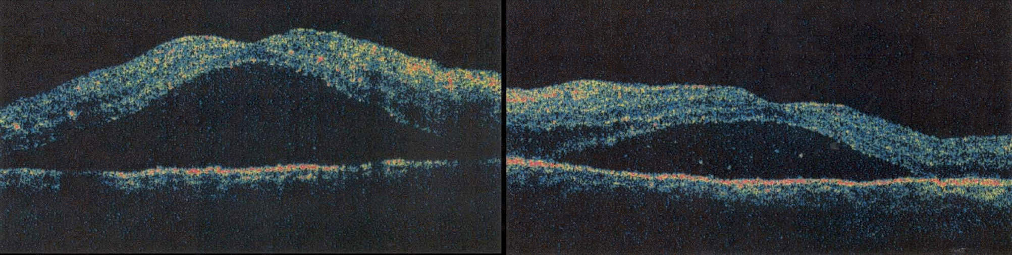
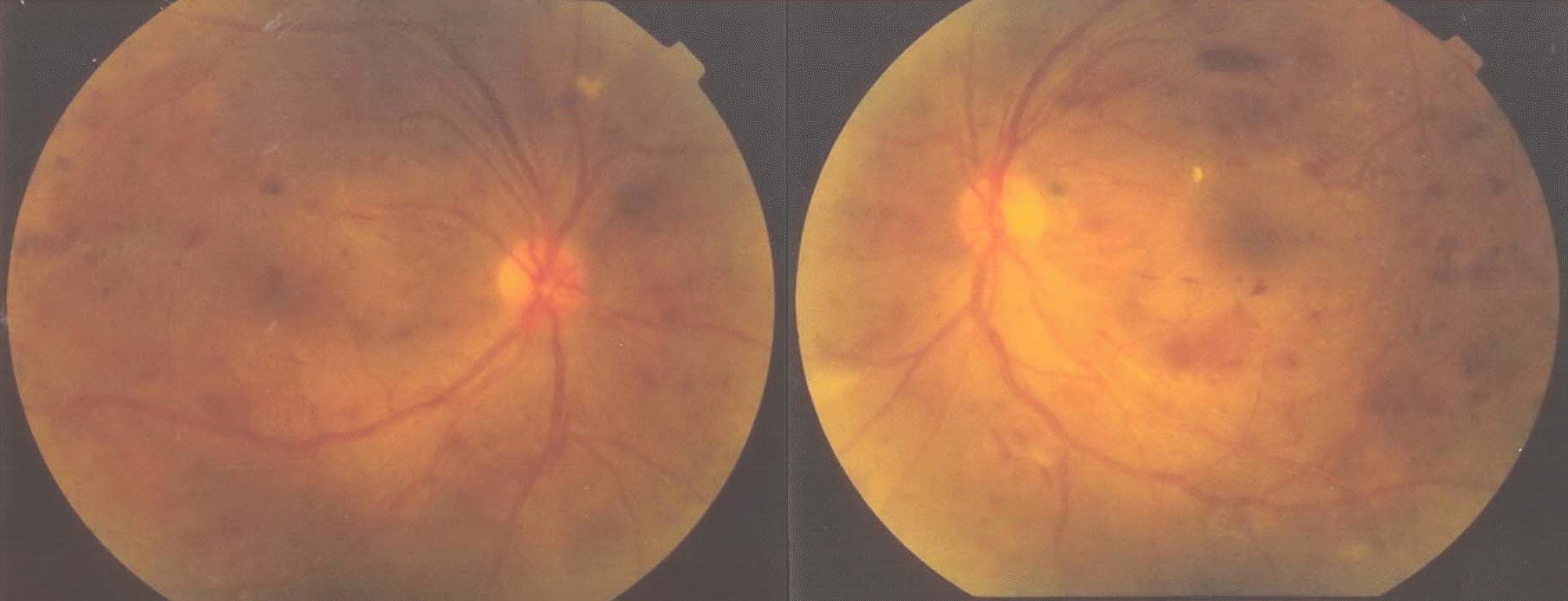
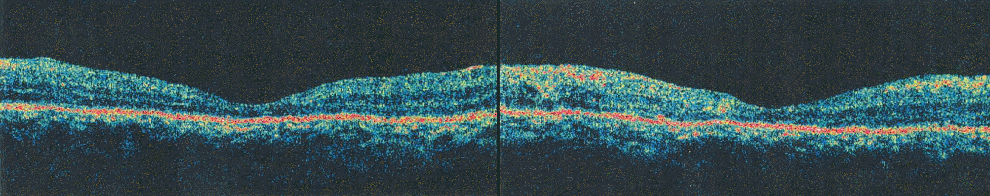
On initial visit, both visual acuity was 0.3. Upon consider changing to fundoscopic examination, the patient was diagnosed with bilateral CRVO. We performed hematologic tests including thrombophilia examination. There were no abnormal findings on routine hematologic tests. Antinuclear antibody and rheumatoid factor were negative but anticardiolipin antibodies presented high titer, on two occasions six weeks apart. We prescribed oral aspirin and performed intravitreal bevacizumab injection under the diagnosis of bilateral CRVO in primary antiphospholipid syndrome.
Go to : 
References
1. Behbehani R, Sergott RC, Savino PJ. The antiphospholipid antibody syndrome: diagnostic aspects. Curr Opin Ophthalmol. 2004; 15:483–5.

2. Miserocchi E, Baltatzis S, Foster CS. Ocular features associated with anticardiolipin antibodies: a descriptive study. Am J Ophthalmol. 2001; 131:451–6.
3. Cobo-Soriano R, Sanchez-Ramon S, Aparicio MJ, et al. Antiphospholipid antibodies and retinal thrombosis in patients without risk factors: a prospective case-control study. Am J Ophthalmol. 1999; 28:725–32.
4. Lahey JM, Tunc M, Kearney J, et al. Laboratory evaluation of hypercoagulable states in patients with central retinal vein occlusion who are less than 56 years of age. Ophthalmology. 2002; 109:126–31.
5. Franchini M, Veneri D. The antiphospholipid syndrome. Hematology. 2005; 10:265–9.
6. Adamczuk YP, Iglesias Varela ML, Martinuzzo ME, et al. Central retinal vein occlusion and thrombophilia risk factors. Blood Coagul Fibrinolysis. 2002; 13:623–6.
7. Castanon C, Amigo MC, Banales JL, et al. Ocular vasoocclusive disease in primary antiphospholipid syndrome. Ophthalmology. 1995; 102:256–62.
8. Miserocchi E, Baltatzis S, Foster CS. Ocular features associated with anticardiolipin antibodies: a descriptive study. Am J Ophthalmol. 2001; 131:451–6.
9. Galetta SL, Plock GL, Kushner MJ, et al. Ocular thrombosis associated with antiphospholipid antibodies. Ann Ophthalmol. 1991; 23:207–12.
10. Al-Abdulla NA, Thompson JT, LaBorwit SE. Simultaneous bilateral central retinal vein occlusion associated with anticardiolipin antibodies in leukemia. Am J Ophthalmol. 2001; 132:266–8.
11. Lahey JM, Tunc M, Kearney J, et al. Laboratory evaluation of hypercoagulable states in patients with central retinal vein occlusion who are less than 56 years of age. Ophthalmology. 2002; 109:126–31.
12. Cobo-Soriano R, Sanchez-Ramon S, Aparicio MJ, et al. Antiphospholipid antibodies and retinal thrombosis in patients without risk factors: a prospective case-control study. Am J Ophthalmol. 1999; 128:725–32.
13. Kim SG, Kim YY, Song GG, et al. Primary antiphospholipid syndrome associated with nonischemic central retinal vein occlusion. J Korean Ophthalmol Soc. 1995; 36:525–30.
14. Kim IT, Na SC, Lee KJ. Vascular Occlusions Associated with Antiphospholipid Antibodies in Systemic Lupus Erythematosus. J Korean Ophthalmol Soc. 2000; 41:427–32.
Go to :
 XML Download