

 PDF
PDF ePub
ePub Citation
 Print
Print


Abstract
Purpose
To report a case of a macular hole formation following rupture of retinal arterial macroaneurysm.
Methods
A 75-year-old female visited our clinic with a chief complaint of decreased vision in her left eye. We completed full ocular examinations, including fluorescein angiography (FAG), and optical coherence tomography (OCT).
Results
The best corrected visual acuity was 0.6 in the right eye and 0.04 in the left. Fundus examination showed a retinal arterial macroaneurysm in supratemporal artery of the left eye. Fluorescein angiography showed hyperfluorescence in the macroaneurysm site. The optical coherence tomography showed macular and submacular hemorrhage. We performed intravitreal gas injection to treat the submacular hemorrhage. One month after the intravitreal gas injection, a full-thickness macular hole developed.
Go to : 
References
1. Flynn HW. Macular hole surgery in patients with proliferative diabetic retinopathy. Arch Ophthalmol. 1994; 112:877–8.

2. Cohen SM, Gass JD. Macular hole following severe hypertensive retinopathy. Arch Ophthalmol. 1994; 112:878–9.
3. Munoz FJ, Rebolleda G, Cores FJ, Bertrand J. Congenital retinal arteriovenous communication associated with a full thickness macular hole. Acta Ophthalmol. 1991; 69:117–20.
4. Rabb MF, Gagliano DA, Teske MP. Retinal arterial macroaneurysms. Surv Ophthalmol. 1988; 33:73–96.
5. Robertson DM. Macroaneurysms of the retinal arteries. Trans Am Acad Ophthalmol Otolaryngol. 1973; 77:55–67.
6. Abdel-Khalek MN, Richardson J. Retinal macroaneurysm: natural history and guidelines for treatment. Br J Ophthalmol. 1986; 70:2–11.
7. Palestin AG, Robertson DM, Goldstein BG. Macroaneurysms of the retinal arteries. Am J Ophthalmol. 1982; 93:164.
8. Cleary PE, Kohner EM, Hamilton AM, et al. Retinal Macroaneurysms. Br J Ophthalmol. 1975; 59:355.
9. Asdourian GK, Goldberg MJ, Jampol L, et al. Retinal macroaneurysms. Arch Ophthalmol. 1977; 95:624–8.
10. Lewis RA, Norton EW, Gass JD. Acquired arterial macroaneurysms of the retina. Br J Ophthalmol. 1976; 60:21–30.
11. Colucciello M, Nachbar JG. Macular hole following ruptured retinal arterial macroaneurysm. Retina. 2000; 20:94–6.
12. Mitamura Y, Terashima H, Takeuchi S. Macular hole formation following rupture of retinal arterial macroaneurysm. Retina. 2002; 22:113–5.
13. Ciardella AP, Barile G, Schiff W, et al. Ruptured retinal arterial macroaneurysm associated with a stage IV macular hole. Am J Ophthalmol. 2003; 135:907–9.
14. Tashimo A, Mitamura Y, Ohtsuka K, et al. Macular hole formation following ruptured retinal arterial macroaneurysm. Am J Ophthalmol. 2003; 135:487–92.
15. Murhty K, Puri P, Talbot JF. Retinal macroaneurysm with macular hole and subretinal neovascular membrane. Eye. 2005; 19:488–9.
Go to :
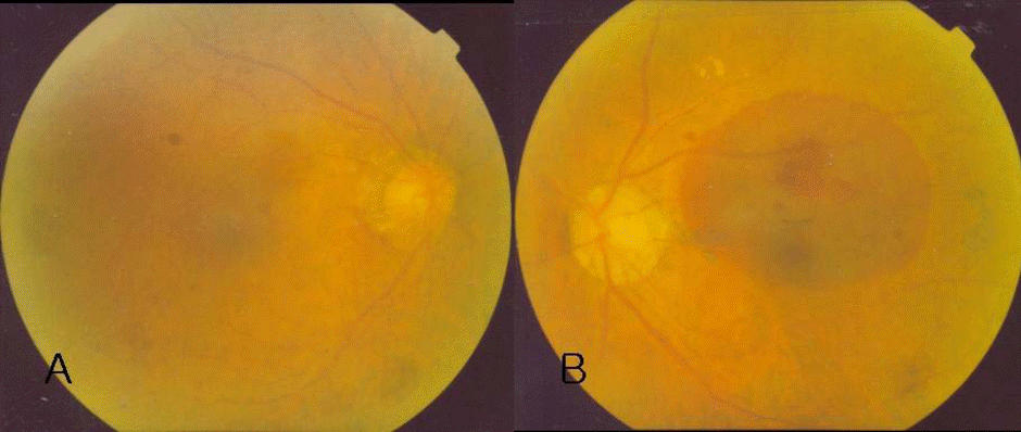 | Figure 1.Fundus photographs. (A) Normal fundus. (B) Subretinal hemorrhage (3-disc diameter area) involving the macular area and macroaneurysm of the yellowish fusiform dilatation lesion (arrow) within the hemorrhage in the superior temporal arcade. |
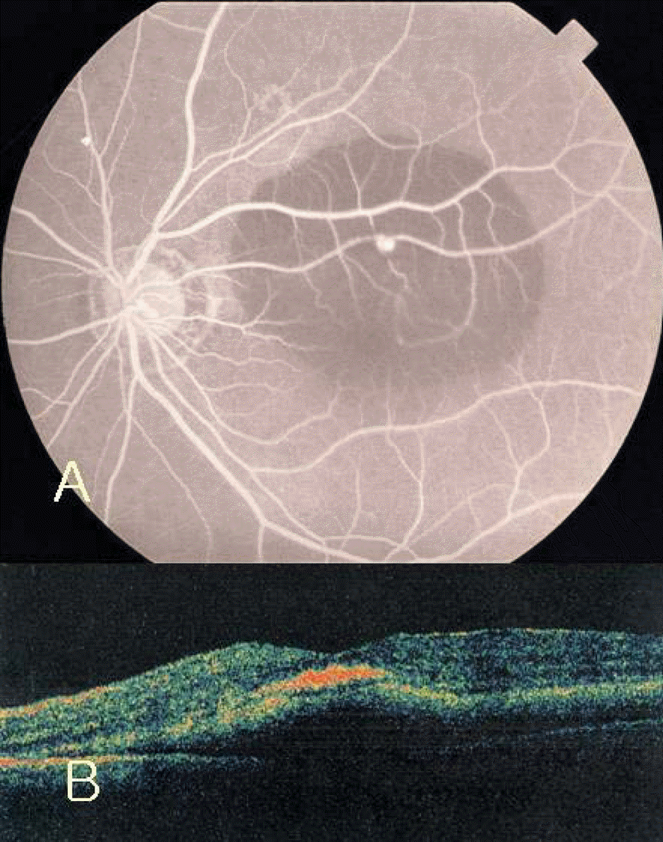 | Figure 2.(A) Fluorescein angiogram (24 seconds) revealed round small hyperfluorescence (arrow) along the superior temporal arteriole corresponding to the yellowish lesion in the fundus photogragh within the hypofluorescence area of subretinal hemorrhage. (B) Optical coherence tomography (OCT) showed retinal and subretinal hemorrhage, macular edema and epiretinal membrane. |
 XML Download