

 PDF
PDF ePub
ePub Citation
 Print
Print


Abstract
Purpose
To report a case of macular edema after the use of the oral hypoglycemic agent rosiglitazone.
Methods
A 43-year-old man, who had diabetic mellitus and was on oral rosiglitazone therapy, complained of a visual disturbance in his left eye. After fundus examination and optical coherence tomography, macular edema was observed, therefore rosiglitazone therapy was discontinued.
References
1. Stumvoll M, Hfaring HU. Glitazones: clinical effects and molecular mechanisms. Ann Med. 2002; 34:217–24.

2. Mudaliar S, Chang AR, Henry RR. Thiazolidinediones, peripheral edema, and type 2 diabetes: incidence, pathophysiology, and clinical implications. Endocr Pract. 2003; 9:406–16.
3. Niemeyer NV, Janney LM. Thiazolidinedione-induced edema. Pharmacotherapy. 2002; 22:924–9.
4. Kennedy F. Do thiazolidinediones cause congestive heart failure? Mayo Clin Proc. 2003; 78:1088–91.
5. Kermani A, Garg A. Thiazolidinedione-associated congestive heart failure and pulmonary edema. Mayo Clin Proc. 2003; 78:1088–91.
6. Wang CH, Weisel RD, Liu PP, et al. Glitazones and heart failure: critical appraisal for the clinician. Circulation. 2003; 107:1350–4.
7. Coluciello M. Vision loss due to macular edema induced by rosiglitazone treatment of diabetes mellitus. Arch Ophthalmol. 2005; 123:1273–5.
8. Kendall C, Wooltorton E. Rosiglitazone (Avandia) and macular edema. CMAJ. 2006; 174:623.
9. Ryan EH, Han DP, Ramsay RC, et al. Diabetic macular edema associated with glitazone use. Retina. 2006; 26:562–70.
10. Graham DJ, Green L, Senior JR, et al. Troglitazone induced liver failure: a case study. Am J Med. 2003; 114:299–306.
11. Bresnick GH. Diabetic macular edema. A review. Ophthalmology. 1986; 93:989–97.
12. Perkovich BT, Meyers SM. Systemic factors affecting diabetic macular edema. Am J Ophthalmol. 1988; 105:211–2.
13. Nguyen QD, Tatlipinar S, Shah SM, et al. Vascular endothelial growth factor is a critical stimulus for diabetic macular edema. Am J Ophthalmol. 2006; 142:961–9.
Figure 1.
Optical coherence tomography (OCT) finding of the patient's left eye, 1 month after the development of visual disturbance showed cyst-like accumulation of subretinal fluid at the central macula.
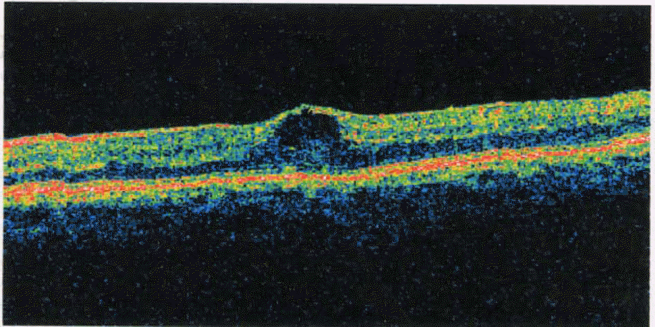
Figure 2.
Optical coherence tomography (OCT) of the patient's left eye, two weeks after discontinuation of rosiglitazone showed marked decrease of subretinal fluid at the central macula.
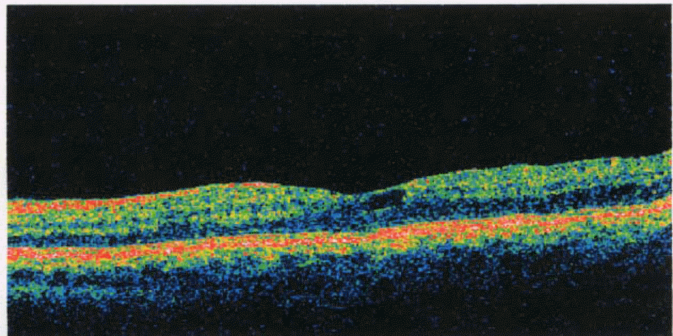
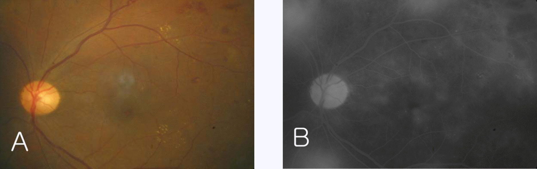
Figure 3.
(A) Fundus photo of the patient‘s left eye, two weeks after discontinuation of rosiglitazone showed multiple dot and blot shaped retinal hemorrhages and circinate hard exudates around macula. (B) Late phase fluorescein angiogram showed diffuse fluorescein leaking from new vessel elsewhere and fluorescein pooling at remnant macular edema.
 XML Download