

 PDF
PDF ePub
ePub Citation
 Print
Print


Abstract
Purpose
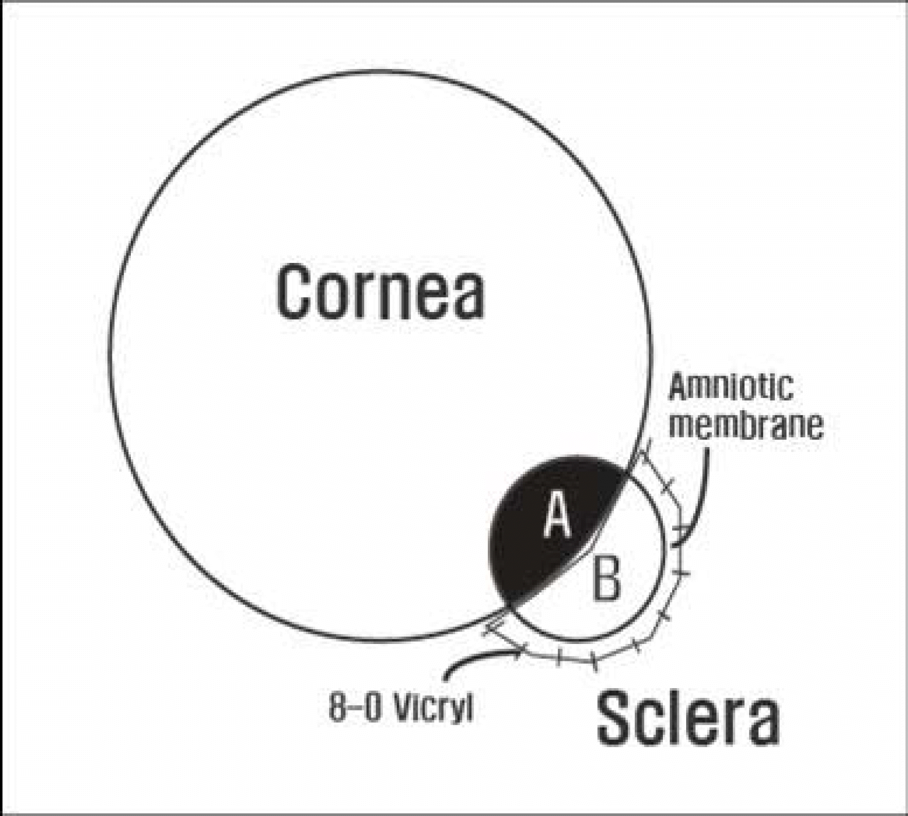
To report two cases of cosmetic treatment of limbal dermoid, which consist of local excision of the limbal dermoid, corneal tattooing, and amniotic membrane transplantation.
Methods
Dermoid excision, corneal tattooing, and amniotic membrane transplantation were carried out in both a girl and a woman who each had a limbal dermoid in their left eyes. Postoperatively, they were followed up for more than six months.
Go to : 
References
1. Mansour AM, Barber JC, Reinecke RD, Wang FM. Ocular choristomas. Surv Ophthalmol. 1989; 33:339–58.

2. Spencer WH. Ophthalmic pathology: an atlas and textbook. 4th ed.4. Philadelphia: W. B. Saunders;1996. p. 50.
3. Scott JA, Tan DT. Therapeutic lamellar keratoplasty for limbal dermoids. Ophthalmology. 2001; 108:1858–67.
4. Park BI. Three cases of conjunctival dermoid. J Korean Ophthalmol Soc. 1962; 3:49–51.
5. Panton RW, Sugar J. Excision of limbal dermoids. Ophthalmic Surg. 1991; 22:85–9.
6. Kim HB, Kim SD, Lew HM, Kwon JS. Two cases of limbal dermoids. J Korean Ophthalmol Soc. 1976; 17:223–7.
7. Kang JH, Park SS. A case of limbal dermoid. J Korean Ophthalmol Soc. 1980; 21:333–6.
8. Hong JS, Jeong CK, Lee TH, Lee HY. A case of limbal complex choristoma containing cartilage. J Korean Ophthalmol Soc. 1998; 39:2187–90.
9. Kim JW, Jin SJ, Rah SH, Cho MY. Osseous choristoma within an epibulbar complex choristoma. J Korean Ophthalmol Soc. 1997; 38:1480–5.
10. Vrabec MP, Jordan JJ, Lawlor PP. Lamellar keratoplasty performed with a corneal scleral button. Ophthalmic Surg. 1994; 25:389–91.
11. Ahn K, Chung ES. Lamellar sclerokeratoplasty for limbal dermoid. J Korean Ophthalmol Soc. 2002; 43:1869–75.
12. Yoon KC, Park SW, Song BY. The role of ultrasound biomicroscopy in operation for limbal dermoid. J Korean Ophthalmol Soc. 2004; 45:364–9.
13. Watts P, Michaeli-Cohen A, Abdolell M, Rootman D. Outcome of lamellar keratoplasty for limbal dermoids in children. J AAPOS. 2002; 6:209–15.
14. Kim C, Han YK, Wee WR, et al. Cosmetic repair of corneal opacity by tattooing. J Korean Ophthalmol Soc. 2005; 46:1967–73.
15. Shen YD, Chen WL, Wang IJ, et al. Full-thickness central corneal grafts in lamellar keratoscleroplasty to treat limbal dermoids. Ophthalmology. 2005; 112:1955–62.
16. Gomes JA, Romano A, Santos MS, Dua HS. Amniotic membrane use in ophthalmology. Curr Opin Ophthalmol. 2005; 16:233–40.
Go to :
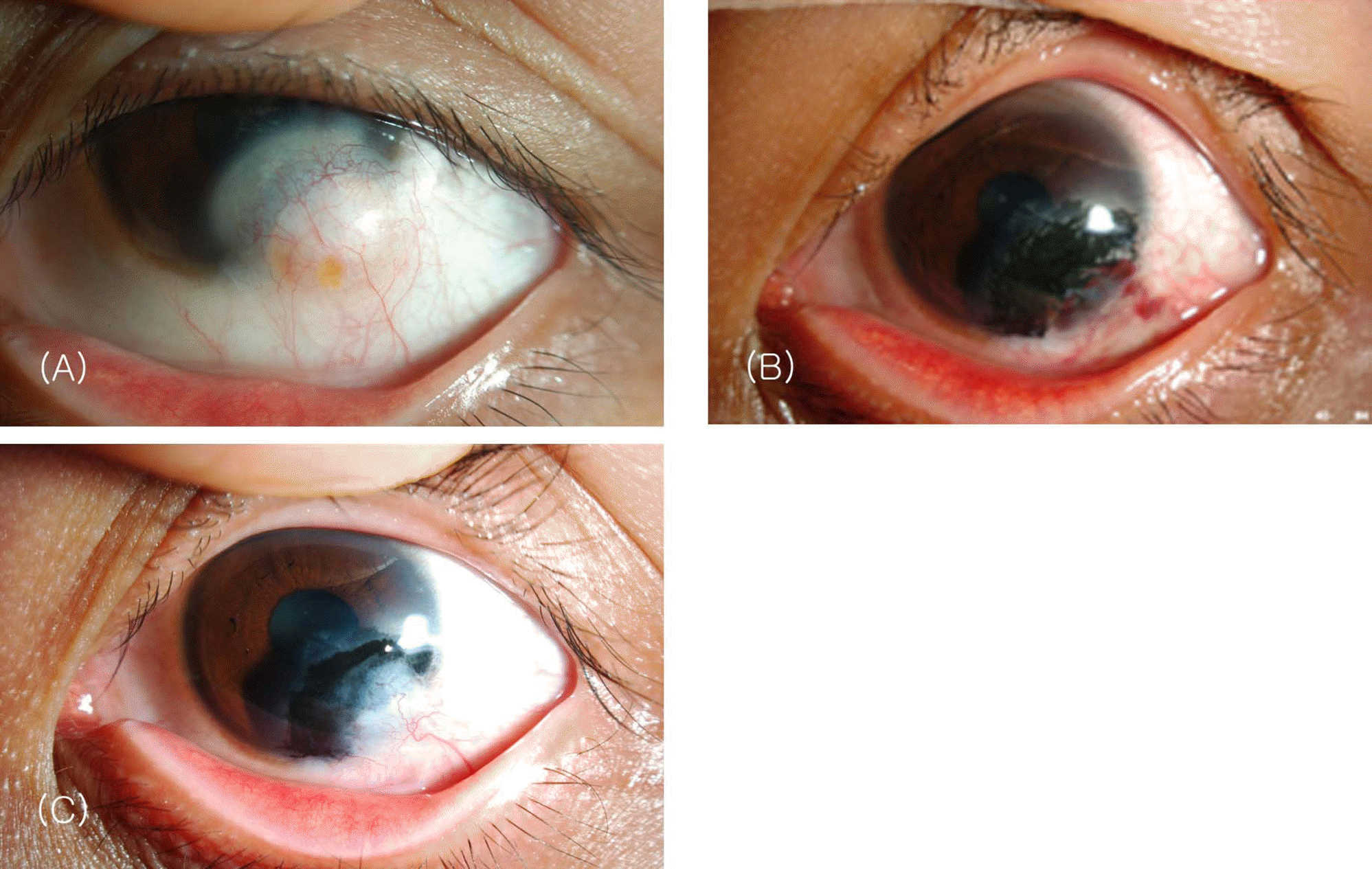
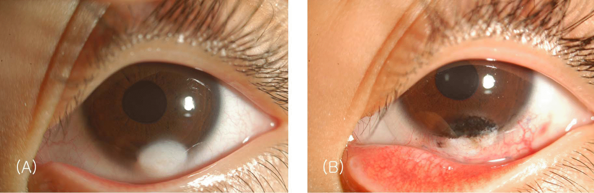 | Figure 1.(A) Limbal dermoid of a 6-year-old girl. (B) Photograph taken at 1 month after limbal dermoid excision, corneal tattooing, and amniotic membrane transplantation. |
 XML Download