

 PDF
PDF ePub
ePub Citation
 Print
Print


Abstract
Purpose
We evaluated the concordance of laterality of the paretic eye and the torsional eye in unilateral superior oblique palsy showing an inferior oblique overaction.
Methods
Thirty-nine patients diagnosed as having a unilateral superior oblique palsy were evaluated for visual acuity, refractive manifestation, ocular movement, prism cover test, and fundus photograph. Of these patients, 32 derived from congenital causes and 7 acquired the condition from trauma. An ocular movement exam was performed to check an inferior oblique overaction, and a fundus photograph was used to measure the ocular torsional amount. Inferior oblique myectomy or recession was performed along with horizontal strabismus surgery.
Results
Objective extorsion was presented in paretic eyes of 29 patients (74.4%) and nonparetic eyes of 10 patients (25.6%). The congenital superior oblique palsy patients were divided into two groups by the concordance of laterality of paretic eyes and torsional eyes. In the concordance group of 22 patients, the torsional amount was decreased from +17.69°to +7.98°and inferior oblique overaction from +2.27°to +0.25°after an inferior oblique muscle weakening procedure. In the discordance group of 10 patients, torsional amount was decreased from +16.97°to +8.73°and inferior oblique overaction from +2.50°to +0.21°postoperatively. In acquired oblique palsy patients, all patients showed the concordance of laterality, and the torsional amount was decreased from +16.76°to +8.80°and inferior oblique overaction from +2.5° to +0.21°after inferior oblique weakening procedure.
Conclusions
We found that the paretic eye and the torsional eye may not coincide in congenital superior oblique palsy but always coincide in acquired oblique palsy after trauma. After an inferior oblique muscle weakening procedure, ocular torsional amount of paretic or sound eye is decreased in every case.
References
1. Von Noorden GK, Campos EC. Binocular vision and ocular Motility. 6th ed.Philadelphia: CV Mosby;1973. p. 177.
2. Von Noorden GK. Clinical observations in cyclodeviations. Ophthalmology. 1979; 86:1451–61.

3. Katherine J, Paul H. Strabismus with a twist: pre- and postoperative torsion. Am Orthopt J. 2003; 53:12–9.
4. Dieterich M, Brandt T. Ocular torsion and perceived vertical in oculomotor, trochlear and abducens nerve palsies. Brain. 1993; 116:1095–104.
5. Sullivan MJ, Kertesz AE. Peripheral stimulation and human cyclofusional response. Invest Ophthalmol Vis Sci. 1979; 18:1287–91.
6. Ruttum M, von Noorden GK. Adaptation to tilting of the visual environment in cyclotropia. Am J Ophthalmol. 1983; 96:229–37.
7. Ruttum M, von Noorden GK. The Bagolini striated lens test for cyclotropia. Doc Ophthalmol. 1984; 58:131–9.
8. Kwon HG, Lee SY, Lee YC. Superior oblique palsy combined with horizontal strabismus. J Korean Ophthalmol Soc. 2003; 44:1846–51.
9. Bixenman WW, von Noorden GK. Apparent foveal displacement in normal subjects and in cyclotropia. Ophthalmology. 1982; 89:58–62.
10. Morton GV, Lucchese N, Kushner BJ. The role of funduscopy and fundus photography in strabismus diagnosis. Ophthalmology. 1983; 90:1186–91.
11. Trobe JD. Cyclodeviation in acquired vertical strabismus. Arch Ophthalmol. 1984; 102:717–20.
12. Metz HS, Norris A. Cyclotorsional diplopia following retinal detachment surgery. J Pediatr Ophthalmol Strabismus. 1987; 24:287–90.
13. Olivier P, von Noorden GK. Excyclotropia of the nonparetic eye in unilateral superior oblique muscle paralysis. Am J Ophthalmol. 1982; 93:30–3.
14. Von Graefe A. Ueber die ophthalmoskospische Beobachtung gewisser Augenmuskelwirkungen. Graefes Arch Ophthalmol. 1856; 2:322.
15. Cho HH, Sohn MA. The ocular cyclotorsion induced by the positional change. J Korean Ophthalmol Soc. 1999; 40:2911–7.
16. Sim JH, Lee SY. The effect of inferior oblique weakening procedures on the correction of ocular torsion. J Korean Ophthalmol Soc. 2005; 46:1020–6.
17. Richard A, Eric L. Abnormal head posture in patients with fourth cranial nerve palsy. Am Orthopt J. 1995; 45:24–33.
18. Kim EH, Lee SJ, Choi HY. Ocular torsion according to fixation in fundus photograph. J Korean Ophthalmol Soc. 2006; 47:449–54.
19. Yim JH, Min BM, Xu YG. Surgical results of classic Harada-Ito procedure with intraoperative adjustment for excyclotorsion. J Korean Ophthalmol Soc. 2002; 43:2227–33.
20. Ohtsuki H, Hasebe S, Hanabusa K, et al. Intraoperative adjustable suture surgery for bilateral superior oblique palsy. Ophthalmology. 1994; 101:188–93.
21. Lee DH, Lee SJ, Park SH. Ocular torsion in normal Korean population. J Korean Ophthalmol Soc. 2004; 45:797–802.
22. Park SW. The torsional status of normal Koreans. J Korean Ophthalmol Soc. 2004; 45:1906–11.
23. Lee HJ, Lim KH. The range of ocular torsion in mass screening. J Korean Ophthalmol Soc. 2005; 46:1684–9.
Figure 1.
The relationships of the inferior oblique overaction and torsional amount in superior oblique palsy. The graph shows a positive correlationship in congenital superior oblique palsy (A). However, in acquired oblique palsy, it is difficult to define correlation because of small number of patients (B).
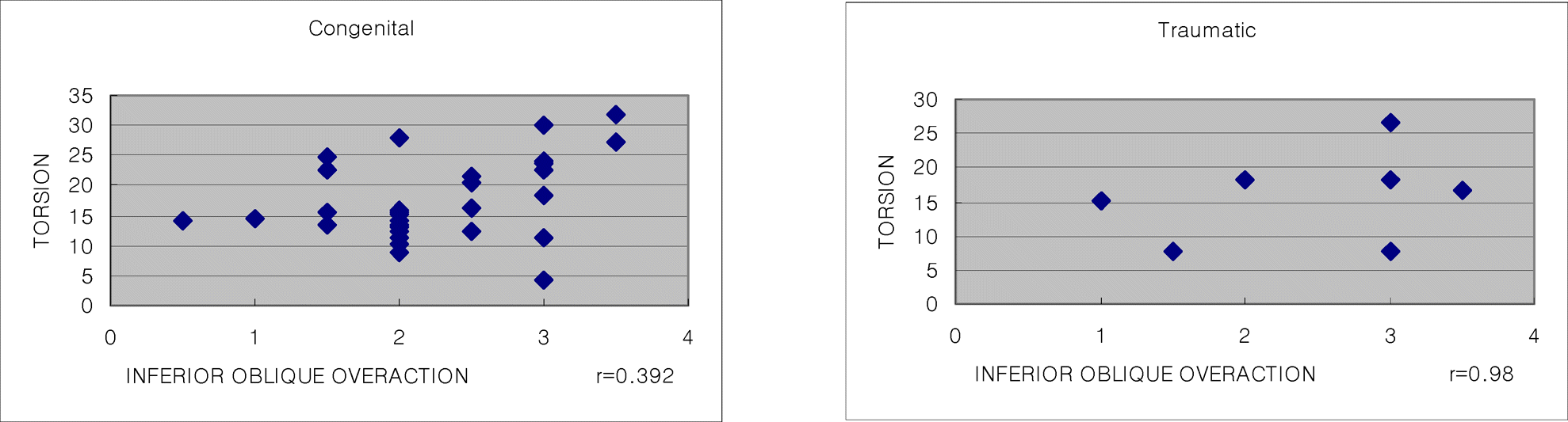
Table 1.
Demographics of patients
| Number of Patients | 39 |
| Average age (years) | 6.4 |
| Sex (M/F) | |
| M | 22 (56.4%) |
| F | 17 (43.6%) |
| SO* palsy | |
| Congenital | 32 (82.1%) |
| Acquired | 7 (17.9%) |
| Weakening procedure | |
| Myectomy | 17 (43.6%) |
| Recession & transposition | 22 (56.4%) |
| Combined with horizontal strabismus | 35 (89.7%) |
| Not combined with horizontal strabismus | 4 (10.3%) |
| Head tilt | 36 (92.3%) |
| Facial asymmetry | 24 (61.5%) |
Table 2.
Changes of the torsional angle in superior oblique palsy
|
Torsion (Op* eye) |
Torsion (TOE†) |
||||
|---|---|---|---|---|---|
| Pre | Post | Pre | Post | ||
| SO‡ palsy | |||||
| Congenital (n=32) | Group 1 (n=22) | 17.69±5.94 | 7.98±6.30 (p=0.0000) | 8.51±4.60 | 8.88±3.98 (p=0.749) |
| Group 2 (n=10) | 10.59±2.73 | 8.09±1.40 (p=0.537) | 16.97±5.09 | 8.73±3.14 (p=0.000) | |
| Acquired (n=7) | Group 1 (n=7) | 16.76±1.39 | 8.80±3.31 (p=0.006) | 9.22±2.94 | 7.02±2.48 (p=0.072) |
| Group 2 (n=0) | - | - | - | - | |
 XML Download