

 PDF
PDF ePub
ePub Citation
 Print
Print


Abstract
Purpose
To analyze peripapillary retinal nerve fiber layer (RNFL) thickness and optic nerve head (ONH) parameters with regard to age in children by using optical coherence tomography (OCT).
Methods
We analyzed RNFL thickness and ONH parameters by using Stratus OCT Model 3000 (Zeiss-Humphrey) in two-hundred eyes of 100 children ranging in age from 5 to 14 years, with 5 males and 5 females for each age.
Results
The RNFL thicknesses for 100 children (200 eyes) in total were as follows: Mean 104.67±9.07 pm, superior 131.84±18.71 pm, temporal 78.49±12.91 pm, nasal 73.85±14.26 pm, and Inferior 133.87±18.58 pm. The parameters of ONH for 100 children (200 eyes) in total were as follows: optic disc area 2.80±0.21 mm2, rim area 1.85±0.28 mm2, cup to disc area ratio 0.34±0.08, horizontal cup to disc diameter ratio 0.47± 0.11, and vertical cup to disc diameter ratio 0.49±0.11. In analyzed RNFL thickness and ONH according to age, there was a significant negative correlation among mean RNFL, inferior RNFL, and age (r=-0.258, p=0.000, r=-0.464, p=0.000). There was a significant positive correlation between nasal RNFL and age (r=0.135, p=0.028). There was a significant positive correlation between disc area and age (r=0.528, p=0.000). There was a significant negative correlation among the cup to disc area ratio, the horizontal cup to disc diameter ratio, the vertical cup to disc diameter ratio, and age (r=-0.170, p=0.008, r=-0.266, p=0.000, r=-0.155, p=0.014).
Go to : 
References
1. Cense B, Chen TC, Park BH, et al. Thickness and Birefringence of Healthy Retinal Nerve Fiber Layer Tissue Measured with Polarization-Sensitive Optical Coherence Tomography. Invest Ophthalmol Vis Sci. 2004; 45:2606–12.

2. Schuman JS, Hee MR, Puliafito CA, et al. Quantification of nerve fiber layer thickness in normal and glaucomatous eyes using optical coherence tomography. Arch Ophthalmol. 1995; 113:586–96.
3. Yamada H, Yamakawa Y, Chiba M, Wakakura M. Evaluation of the effect of aging on retinal nerve fiber thickness of normal Japanese measured by optical coherence tomography. Nippon Ganka Gakkai Zasshi. 2006; 110:165–70.
4. Kanamori A, Escano MF, Eno A, et al. Evaluation of the effect of aging on retinal nerve fiber layer thickness measured by optical coherence tomography. Ophthalmology. 2003; 217:273–8.
5. Chi Q, Goji T, Yoskiaki K. Evaluation of the effect of aging on the retinal nerve fiber layer thickness using scanning laser polarimetry. Zhonghua Yan Ke Za Zhi. 1998; 34:199–201.
6. Park SE, Choi KR. Analysis of optic disc size and retinal nerve fiber thickness. J Korean Ophthalmol Soc. 2002; 43:395–401.
7. Mendez MS, Gonzalez-Hernandez M, Lozano-Lopez V, et al. Optic disc tomography and perimetry in controls, glaucoma suspects, and early and established glaucomas. Optom Vis Sci. 2007; 84:33–41.
8. Skorkovska K, Skorkovska S, Michalek J, Koci J. Influrence of age, gender, refraction, Keratometry and disc area on the topographic parameters of the optic nerve head. Cesk Slov Oftalmol. 2005; 61:245–52.
9. Sanchez-Galeana C, Bowd C, Blumenthal EZ, et al. Using optical imaging summary data to detect glaucoma. Ophthalmology. 2001; 108:1812–8.
10. Pueyo V, Polo V, Larrosa JM, et al. Reproducibility of optic nerve head and retinal nerve fiber layer thickness measurements using optical coherence tomography. Arch Soc Esp Ofthalmol. 2006; 81:205–11.
11. Kamppeter BA, Schubert KV, Budde WM, et al. Optical coherence tomography of the optic nerve head: interindividual reproducibility. J Glaucoma. 2006; 15:248–54.
12. Kanamori A, Nakamura M, Escano MF, et al. Evaluation of the glaucomatous damage on retinal nerve fiber layer thickness measured by optical coherence tomography. Am J Ophthalmol. 2003; 135:513–20.
13. Ozdek SC, Onol M, Gurelik G, Hasanreisoglu B. Scanning laser polarimetry in normal subjects and patients with myopia. Br J Ophthalmol. 2000; 84:264–7.
14. Bowd C, Zangwill LM, Blumenthal EZ, et al. Imaging of the optic disc and retinal nerve fiber layer: The effects of age, optic disc area, refractive error, and gender. J Opt Soc Am A Opt Image Sci Vis. 2002; 19:197–207.
15. Oliveira C, Harizman N, Girkin CA, et al. Axial length and optic disc size in normal eyes. Br J Ophthalmol. 2007; 91:37–9.
16. Altintas O, Yuksel N, Ozkan B, Caglar Y. Thickness of the retinal nerve fiber layer, macular thickness, and macular volume in patients with strabismic amblyopia. J Pediatric Ophthalmol Strabismus. 2005; 42:216–21.
17. Reche-Sainz JA, Domingo-Gordo B, Toledano-Femandez N. Study of the retinal nerve fiber layer in childhood strabismus. Arch Soc Esp Ofthalmol. 2006; 81:21–5.
18. Huynh SC, Wang XY, Rochtchina E, et al. Distribution of optic dise parameters measured by OCT: findings from a population-based study of 6-year-old Australian dhildren. Invest Ophthalmol Vis Sci. 2006; 47:3276–85.
19. Huynh SC, Wang XY, Rochtchina E, et al. Peripapillary retinal nerve fiber layer thickness in a population of 6 year-old children: finding by optical coherence tomography. Ophthalmology. 2006; 113:1583–92.
20. Saichow DJ, Oleynikov YS, Chiang MF, et al. Retinal nerve fiber layer thickness in normal children measured with optical coherence tomography. Ophthalmology. 2006; 113:786–91.
21. Ahn HC, Son HW, Kim JS, Lee JH. Quantification analysis of retinal nerve fiber layer thickness of normal children and adolescents. Korean J Ophthalmol. 2005; 19:195–200.
22. Choi MG, Han M, Kim YI, Lee JH. Comparison of glaucomatous parameters in normal, ocular hypertensive and glaucomatous eyes using optical coherence tomography 3000. Korean J Ophthalmol. 2005; 19:40–6.
23. Baquerp aranda IM, Morillo sanchez MJ, Garcia campos JM. Use of optical coherence tomography to study variations of normal parameter with age. Arch Soc Esp Oftalmol. 2005; 80:225–31.
Go to :
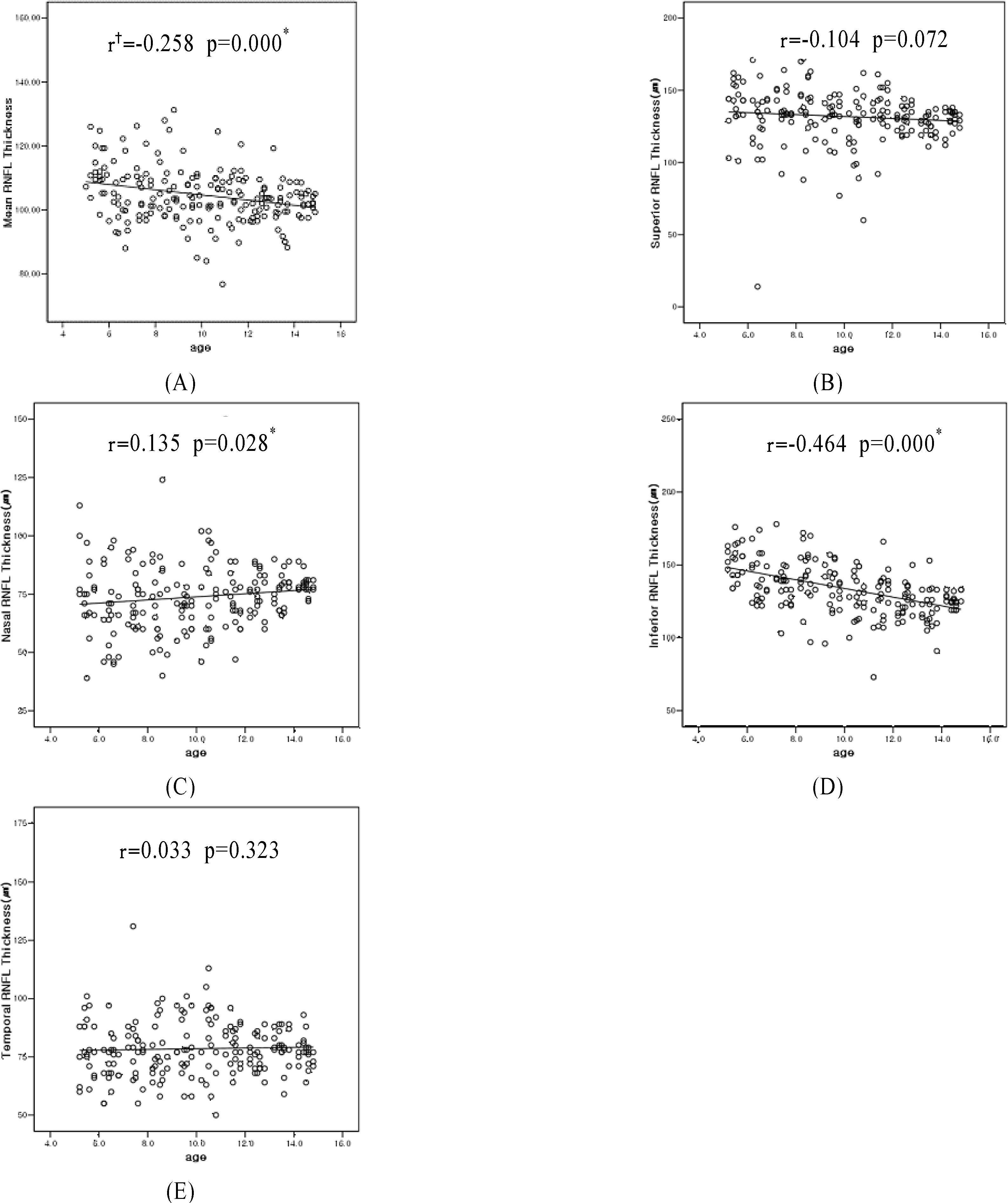 | Figure 1.Retinal nerve fiber layer (RNFL) thickness (pm) in children by age (200 eyes). (A) Mean RNFL thickness, (B) Superior RNFL thickness, (C) Nasal RNFL thickness, (D) Inferior RNFL thickness, (E),Temporal RNFL thickness. Correlation coefficient. |
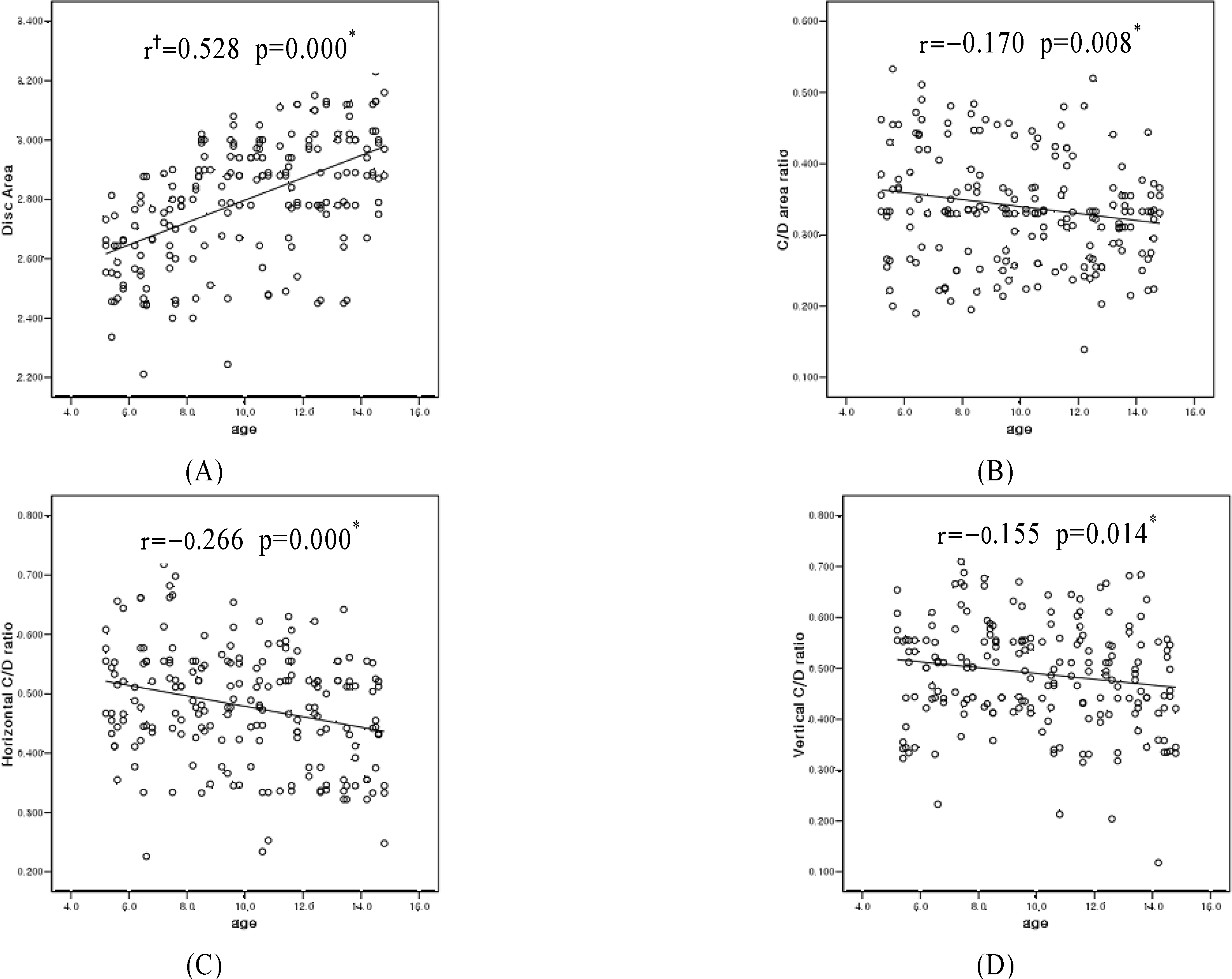 | Figure 2.Optic nerve head parameter in children by age (200 eyes). (A) Disc Area, (B) C/D* area ratio, (C) Horizontal C/D ratio, (D) Vertical C/D ratio (D). Cup to Disc. |
Table 1.
Study characteristics
| Age (year) | 5 | 6 | 7 | 8 | 9 | 10 | 11 | 12 | 13 | 14 |
|---|---|---|---|---|---|---|---|---|---|---|
| Gender | 5:5 | 5:5 | 5:5 | 5:5 | 5:5 | 5:5 | 5:5 | 5:5 | 5:5 | 5:5 |
| (Male:Female) (No *) | ||||||||||
| Total eyes (No *) | 20 | 20 | 20 | 20 | 20 | 20 | 20 | 20 | 20 | 20 |
| Mean IOP† ±SD‡ | 14.35 | 15.90 | 15.55 | 16.90 | 14.65 | 14.70 | 13.80 | 15.05 | 15.40 | 15.10 |
| (mmHg) | ±2.72 | ±2.25 | ±2.56 | ±1.68 | ±2.11 | ±2.03 | ±1.64 | ±1.85 | ±1.82 | ±1.52 |
| Mean P§±SD | +0.31 | +0.35 | +0.55 | -0.25 | -1.25 | -0.25 | -0.48 | -1.25 | -0.45 | -0.30 |
| (Diopter) | ±1.53 | ±1.37 | ±1.22 | ±1.24 | ±1.36 | ±1.31 | ±1.48 | ±1.31 | ±1.25 | ±1.19 |
Table 2.
Analysis of retinal nerve fiber layer (RNFL) thickness in 100 children (200 eyes).
| RNFL (μm) | Mean±SD∗ |
|---|---|
| Mean | 104.67±9.07 |
| Superior | 131.84±18.71 |
| Temporal | 78.49± 12.91 |
| Nasal | 73.85± 14.26 |
| Inferior | 133.87± 18.58 |
Table 3.
Analysis of optic nerve head parameter in 100 children (200 eyes)
| Mean±SD* | |
|---|---|
| Disc Area (mm2) | 2.80±0.21 |
| Rim Area (mm2) | 1.85±0.28 |
| C/D† area ratio | 0.34±0.08 |
| Horizontal C/D ratio | 0.47±0.11 |
| Vertical C/D ratio | 0.49±0.11 |
Table 4.
Analysis of retinal nerve fiber layer (RNFL) thickness according to age
| Age (year) | Mean | Superior | Mean±SD* (μm) Temporal | Nasal | Inferior |
|---|---|---|---|---|---|
| 5 | 112.08±6.98 | 140.30± 16.45 | 79.10±12.49 | 76.10±15.88 | 152.55±11.58 |
| 6 | 102.88±9.05 | 125.80±31.64 | 72.30± 10.09 | 66.75±16.64 | 140.35±15.76 |
| 7 | 106.13±7.89 | 135.50±15.26 | 79.30±15.34 | 73.60±11.41 | 136.10±14.90 |
| 8 | 108.01±10.33 | 139.00±20.92 | 76.45± 12.33 | 71.55±19.89 | 143.25±18.69 |
| 9 | 103.30±7.94 | 129.70±17.01 | 79.40± 12.85 | 68.85±8.13 | 135.25±15.28 |
| 10 | 104.85±15.74 | 122.20±23.56 | 86.40±24.22 | 76.00±17.59 | 134.80±28.31 |
| 11 | 104.41±7.50 | 136.50±15.39 | 80.00±8.42 | 75.00±20.47 | 124.15± 19.52 |
| 12 | 101.68±3.84 | 130.75±8.14 | 75.45±6.96 | 75.85±8.67 | 124.65±10.08 |
| 13 | 100.56±7.59 | 128.50±14.07 | 79.60±7.78 | 76.20±7.58 | 120.00±13.50 |
| 14 | 102.81±3.28 | 130.10±7.23 | 76.90±6.43 | 78.65±4.18 | 125.60±4.69 |
| F† (p-value) | 3.041 (0.002)‡ | 2.045 (0.057) | 1.678 (0.097) | 1.382 (0.199) | 7.405(0.000)‡ |
Table 5.
Analysis of optic nerve head parameter according to age
| Age (year) | C/D† area ratio | Mean±SD∗ Horizontal C/D ratio | Vertical C/D ratio |
|---|---|---|---|
| 5 | 0.354±0.086 | 0.504±0.081 | 0.475±0.107 |
| 6 | 0.397±0.104 | 0.484±0.106 | 0.488±0.104 |
| 7 | 0.319±0.083 | 0.550±0.101 | 0.537±0.106 |
| 8 | 0.357±0.081 | 0.479±0.072 | 0.520±0.088 |
| 9 | 0.319±0.073 | 0.493±0.090 | 0.513±0.078 |
| 10 | 0.330±0.061 | 0.453±0.103 | 0.475±0.129 |
| 11 | 0.382±0.097 | 0.500±0.091 | 0.499±0.100 |
| 12 | 0.293±0.086 | 0.447±0.081 | 0.472±0.112 |
| 13 | 0.328±0.047 | 0.456±0.097 | 0.502±0.097 |
| 14 | 0.320±0.055 | 0.424±0.087 | 0.417±0.107 |
| F‡ | 3.282 | 3.062 | 2.070 |
| (p-value) | (0.001)§ | (0.002)§ | (0.034)§ |
 XML Download