Abstract
Methods
A retrospective study was carried out on 11 eyes in 9 patients who had undergone pars plana vitrectomy for Terson's syndrome from October 2004 to June 2006. The factors assessed were age, gender, presence of hypertension, type of intracranial hemorrhage, preoperative and final visual acuity, time interval from intracranial hemorrhage (ICH) to vitrectomy, and any intraoperative and postoperative complications.
Results
The average age of the subjects and the Interval from ICH to vitrectomy were 43.0±11.0 years and 3.25±3.48 months respectively. Binocular involvement was found in two of the nine patients, and fundus findings were severe vitreous opacity in all cases, while sub-ILM hemorrhage at the posterior pole was seen in five eyes. Intraoperative retinal break was recorded at the 10 o'clock sclerotomy site in five eyes, and four of these five eyes were associated with sub-ILM hemorrhage. One patient underwent a scleral buckling operation four months postoperatively due to rhegmatogenous retinal detachment associated with a retinal tear at the 2 o'clock sclerotomy site. Visual acuity improved in all cases postoperatively, and the final visual acuity was over 0.6 in seven eyes.
Conclusions
We can expect from early surgery a relatively good prognosis of visual acuity and prevention of complications. Due to the possibility of retinal breaks at the sclerotomy sites, we should keep in mind that cautious handling of intraocular instrument and complete removal of vitreous base may be necessary.
Go to : 
References
1. Terson A. De L'hemorrhagie dans le corps vitre au cours de I‘hemorrhgie cerebrale. Clin Ophthalmol. 1990; 6:309–12.
2. Kuhn F, Morris R, Witherspoon CD, Mester V. Terson syndrome. Results of vitrectomy and the significance of vitreous hemorrhage in patients with subarachnoid hemorrhage. Ophthalmology. 1998; 105:1261–3.
3. Schultz PN, Sobol WM, Weineist TA. Long-term visual outcome in Terson syndrome. Ophthalmology. 1991; 98:1814–9.

4. Ritland JS, Syrdalen P, Eide N, et al. Outcome of vitrectomy in patients with Terson syndrome. Acta Ophthalmol Scand. 2002; 80:172–5.

5. Gnanaraj L, Tyagi AK, Cottrell DG, et al. Referral delay and ocular surgical outcome in Terson syndrome. Retina. 2000; 20:374–7.

6. Kim US, Yu SY, Kwak HW. Incidence and postoperative visual outcome of Terson's syndrome. J Korean Ophthalmol Soc. 2002; 43:2451–6.
7. Song TH, Lee ES, Jin HS, et al. Incidence, treatment and prognosis of Terson's Syndrome. J Korean Ophthalmol Soc. 2006; 47:763–70.
8. Yokoi M, Kase M, Hyodo T, et al. Epiretinal membrane formation in Terson syndrome. Jpn J Ophthalmol. 1997; 41:168–73.

10. Wietholter S, Steube D, Stotz HP. Terson syndrome: a frequently missed ophthalmologic complication in subarachnoid hemorrhage. Zentralbl Neurochir. 1998; 59:166–70.
11. Velikay M, Datlinger P, Stolba U, et al. Retinal detachment with severe proliferative vitreoretinopathy in Terson syndrome. Ophthalmology. 1994; 101:35–7.

12. Isernhagen RD, Smiddy WE, Michels RG, et al. Vitrectomy for nondiabetic vitreous hemorrhage. Not associated with vascular disease. Retina. 1988; 8:81–7.
13. Murjaneh S, Hale JE, Mishra S, et al. Terson's syndrome: surgical outcome in relation to entry site pathology. Br J Ophthalmol. 2006; 90:512–3.

14. Miller-Meeks MJ, Bennett SR, Keech RV, Blodi CF. Myopia induced by vitreous hemorrhage. Am J Ophthalmol. 1990; 109:199–203.

15. Clarkson JG, Flynn HW, Daily MJ. Vitrectomy in Terson's syndrome. Am J Ophthalmol. 1980; 90:549–52.

16. Friedman SM, Margo CE. Bilateral subinternal limiting membrane hemorrhage with Terson syndrome. Am J Ophthalmol. 1997; 124:850–1.

17. Garcia-Arumi J, Corcostegui B, Tallada N, Salvador F. Epiretinal membranes in Tersons syndrome. A clinicopathologic study. Retina. 1994; 14:351–5.
Go to : 
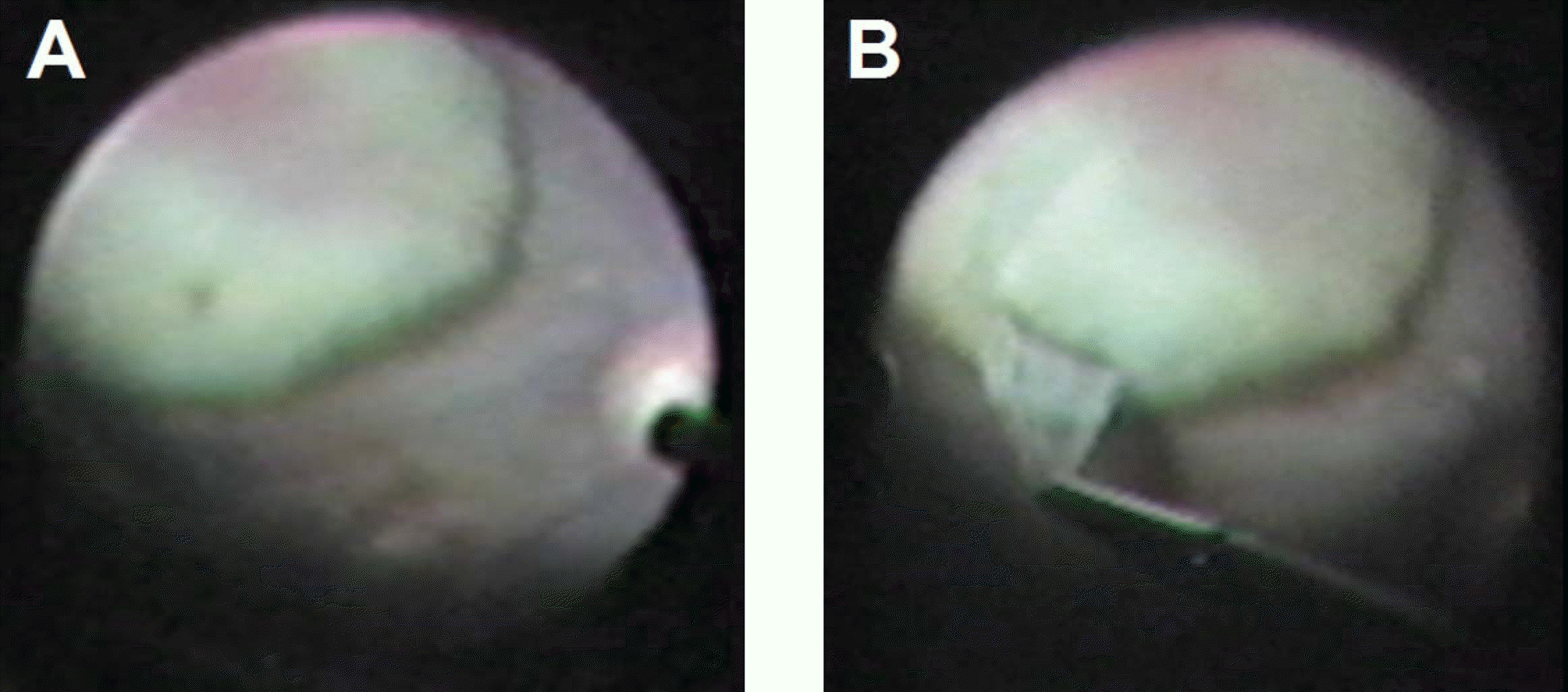
 | Figure 1.(A) The discrete Large dome-shaped subinternal limiting membrane hemorrhage in the posterior pole (B) Internal limiting membrane was peeled off with the microforceps at an area of subinternal limiting membrane hemorrhage using indocyanine green. |
Table 1.
Baseline patient data
Table 2.
Clinical data
Table 3.
Surgical outcome post-vitrectomy for Terson's syndrome and nondiabetic vitreous hemorrhage


 PDF
PDF ePub
ePub Citation
 Print
Print


 XML Download