

 PDF
PDF ePub
ePub Citation
 Print
Print


Abstract
Purpose
To investigate the courses leading to bilateral enucleation in bilateral retinoblastoma patients.
Methods
Medical records of 5 bilateral retinoblastoma patients who underwent bilateral enucleation were reviewed for patient information, history, change of tumor state during the treatment and the cause of bilateral enucleation.
Results
Out of 48 bilateral retinoblastoma patients, both eyes were saved in 6 children, 1 eye was saved in 37 children, and the remaining 5 children lost both eyes. All patients who underwent bilateral enucleation were female and had no family history of retinoblastoma. At diagnosis, 3 children were 4 months old and the remaining 2 patients were 1 year and 1.5 years old each. Out of 10 eyes, 8 eyes were in Reese Ellsworth group V and the remaining 2 eyes were in group III. The initial treatment was enucleation of 1 eye followed by chemotherapy in 3 patients, and chemotherapy alone in 2 patients. Additional treatment included laser photocoagulation, cryotherapy, external beam radiation therapy and proton beam irradiation. Vitreous seeding, development of new tumors, and increase in tumor size despite of intensive, conservative treatment resulted in second enucleation.
Go to : 
References
1. Ellsworth RM. Practical management of retinoblastoma. Trans Am Ophthalmol Soc. 1969; 67:462–534.
2. Pendergrass TW, Davis S. Incidence of retinoblastoma in the UnitedStates. Arch Ophthalmol. 1980; 98:1204–10.
3. Schappert-Kimmijser T, Hemmes GD, Nijland R. The heredity of retinoblastoma. Ophthalmologica. 1996; 151:197.

4. Jensen RD, Miller RW. Retinoblastoma: Epidemiologic characteristics. N Engl J Med. 1971; 285:307.
5. Binder PS. Unusual manifestations of retinoblastoma. Am J Ophthalmol. 1974; 77:674–9.
6. Kincaid MC. Ocular and orbital tumors. Ophthalmology. 1988; 95:1588–95.
7. Shields CL, Shields JA, De Potter P. New treatment modalities for retinoblastoma. Curr Opin Ophthalmol. 1996; 7:20–6.
8. Abramson DH, Niksarli K, Ellsworth RM, et al. Changing trends in the management of retinoblastoma: 1951-1965 vs 1966-1980. J Pediatr Ophthalmol Strabismus. 1994; 31:32–7.
9. Sagerman RH, Cassady R, Tretter P, Ellsworth RM. Radiation induced neoplasia following external beam therapy for children with retinoblastoma. Am J Roentgenol Radium Ther Nucl Med. 1969; 105:529–35.
10. Roarty JD, McLean IW, Zimmerman LE. Incidence of second neoplasia in patients with bilateral retinoblastoma. Ophthalmology. 1984; 95:1583–7.
11. Abramson DH, Ellsworth RM, Kitchin D, Tung G. Second nonocular tumors in retinoblastoma survivors, Are they radiation-induced? Ophthalmology. 1984; 91:1351–5.
12. Seregard S, Lundell G, Svedberg H, Kivela T. Incidence of retinoblastoma from 1958 to 1998 in Northern Europe: advantages of birth cohort analysis. Ophthalmology. 2004; 111:1228–32.
13. Park JH, Chung HK, Khwarg SI, Yu YS. Clinical Course of Spontaneous Regression of Bilateral Retinoblastoma. J Korean Ophthalmol Soc. 2006; 47:2047–52.
14. Sanders BM, Draper GJ, Kingston JE. Retinoblastoma in Great Britain 1969-1980: incidence, treatment and survival. Br J Ophthalmol. 1988; 72:576–83.
15. Lee JB, Shim IC, Oh JS. Clinical Observations of Retinoblastoma. J Korean Ophthalmol Soc. 1982; 23:77–84.
16. Yang JG, Yu YS. Clinical Characteristics of the Retinoblastoma Diagnosed before One Year Old. J Korean Ophthalmol Soc. 1996; 37:85–91.
17. Song JS, Lee JK, Lee TW. Treatment and prognosis of retinoblastoma : Clinicopathologic analysis of 101 cases. J Korean Ophthalmol Soc. 1998; 39:2393–405.
18. Midgal C. Bilateral retinoblastoma:the prognosis for vision. Br J Ophthalmol. 1983; 67:592–5.
19. Lee V, Hungerford JL, Ahmed F, et al. Globe conserving treatment of the only eye on bilateral retinoblastoma. Br J Ophthalmol. 2003; 87:1374–80.
20. Shields JA, Shield CL. Retinoblastoma; Clinical and Pathologic features in Intraocular tumors, A Text and Atlas. 1st ed.1. Philadelphia: WB Sanders;1992. p. 305–32.
21. Abramson DH, Schefler AC. Update on retinoblastoma. Retina. 2004; 24:828–48.
22. Kim JH, Yu YS, Khwarg SI, et al. Clinical Result of Prolonged Primary Chemotherapy in Retinoblastoma Patients. Korean J Ophthalmol. 2003; 17:35–43.
23. Shields CL, Honavar SG, Meadows A, et al. Chemoreduction plus focal therapy for retinoblastoma : factors predictive of need for treatment with external beam radiotherapy or enucleation. Am J Ophthalmol. 2002; 133:657–64.
24. Abramson DH, Notterman RB, Ellsworth RM. Retinoblastoma treated in infants in the first six months of life. Arch Ophthalmol. 1983; 108:1362–7.
25. Lee TW, Yang SW, Kim BH. Clinical Analysis of the Retinoblastoma. J Korean Ophthalmol Soc. 1995; 36:96–105.
26. Eng C, Li FP, Abramson DH, et al. Mortality from second tumours among long-term survivors of retinoblastoma. J Natl Cancer Inst. 1993; 85:1121–8.
27. Winick NJ, Mckenna RW, Shuster JJ, et al. Secondary acute myeloid leukemia in children with acute lymphoblastic leukemia treated with etoposide. J Clin Oncol. 1993; 11:209–17.
28. Abramson DH, Lee TC, Driscoll C. Osteogenic sarcoma in a survivor of metastatic germline retinoblastoma: the possible influence of human growth hormone. J Pediatr Ophthalmol Strabismus. 2002; 39:347–8.
Go to :
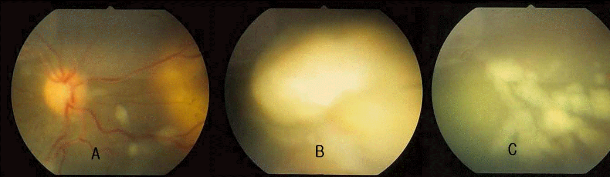 | Figure 2.Fundus photographs of the right eye of case 1. (A): multiple vitreous seedings on the posterior pole and regressed mass at 3 o'clock, (B) large new tumor mass at 2∼3 o'clock, and (C): multiple vitreous seedings in the inferonasal area. |
Table 1.
Characteristics of patients at diagnosis
| Case | Sex/Age | FHx∗ | R/L | Chief Complaint | Time to Dx† |
|---|---|---|---|---|---|
| 1 | F/18 m | no | R | Found on examination | 2 w ‡ |
| L | Conjunctival injection and lid edema | ||||
| 2 | F/4 m | no | R | Leucocoria | 3 m § |
| L | Leucocoria | ||||
| 3 | F/4 m | no | R | Found on examination | 1 w |
| L | Leucocoria | ||||
| 4 | F/4 m∏ | no | R | Esodeviation | 2 w |
| L | Esodeviation | ||||
| 5 | F/12 m | no | R | Poor fixation | 3 mo |
| L | Poor fixation |
Table 2.
Clinical characteristics at the diagnosis
| Case | R/L | Mass on funduscopy | R-E∗ | Calcification | Brain MRI | Bone scan | Prim Tx† |
|---|---|---|---|---|---|---|---|
| 1 | R | 2 (4∼6DD‡, 1.5DD) Vit. seeding (+) | Vb | (+) | (−) | (−) | Chemo§ |
| L | Full vol. of vit. cavity | Va | (+) | En∏ | |||
| 2 | R | Full vol. of vit. cavity | Va | (+) | En | ||
| L | 4 (1∼2DD x 2 (anterior to equator, 4∼5DD, 8DD) | IIIa | (−) | (−) | (−) | Chemo | |
| 3 | R | Larger than 10DD | IIIb | (+) | Chemo, L# | ||
| L | 1/2 vol. of vit cavity | Va | (+) | (−) | (−) | En | |
| 4 | R | Full vol. of vit cavity | Va | Not done | Chemo | ||
| L | Full vol. of vit cavity | Va | Not done | (−) | (−) | Chemo | |
| 5 | R | 1/2 vol. of vit cavity | Va | (+) | Chemo | ||
| L | 1/2 vol. of vit cavity | Va | (+) | (−) | (−) | Chemo |
Table 3.
Treatment course 1
| Case | Chemotherapy (cycles) | Local Tx | RadioTx | Tx response | Outcome | ||
|---|---|---|---|---|---|---|---|
| 1/R | AΠ | A | B | C∗ x2 | EBR§ x22 | Vit seeding ↑ | Enucleation |
| 12 | 7 | 1 | L† x1 | New, multiple | |||
| T‡ x1 | tumors | ||||||
| 2/L | A | L x1 | Vit seeding ↑ | Enucleation | |||
| 6 | New tumors | ||||||
| Size ↑ | |||||||
| 3/R | A | B# | A | C x1 | Size ↑ | Enucleation | |
| 14 | 10 | 4 | L x5 | ||||
| 4/R | A | A | C x1 | Proton | Size ↑ | Enucleation | |
| 13 | 2 | beam | |||||
| 4/L | A | A | Size ↑ | Enucleation | |||
| 13 | 2 | New tumors | |||||
| 5/R | A | B | A | EBR x25 | Size ↑ | Enucleation | |
| 8 | 1 | 10 | |||||
| 5/L | A | B | A | C x1 | EBR x25 | New, multiple | Enucleation |
| 8 | 1 | 10 | L x1 | tumors | |||
Table 4.
Treatment course 2
| Case | Interval btn∗ enucleations | Reason of 2nd eye enucleation | F/U† brain MRI & bone scan | F/U after 2nd enucleation |
|---|---|---|---|---|
| 1 | 22 m | Vit seeding ↑ | (−)‡ | 16 m |
| Multiple new tumors (+) | ||||
| 2 | 8 m | Multiple new tumors (+) | (−) | 19 m |
| Vit seeding ↑ | ||||
| Size ↑ | ||||
| 3 | 55 m | Size ↑ | (−) | 29 m |
| 4 | 14 m | Size ↑ | (−) | 30 m |
| 5 | 8 m | Size ↑ | (−) | 22 m |
 XML Download