

 PDF
PDF ePub
ePub Citation
 Print
Print


Abstract
Purpose
The aim of this study is to evaluate the effects and complications of periocular injections of triamcinolone acetonide in patients with thyroid orbitopathy who could not tolerate systemic corticosteroid therapy.
Methods
Six patients with a mean age of 48.7 years showed symptoms of severe acute thyroid orbitopathy. They received four doses of 20 mg of triamcinolone acetonide via periocular injection into the inferotemporal orbital quadrant every 2 weeks. The response to treatment and the presence of adverse effects were evaluated retrospectively.
Results
Three of six patients (50%) showed significant improvement in soft tissue swelling in both eyes. Only one patient (17%) showed improvement of proptosis. No patients showed improvement in diplopia and ocular motility. The mean thickness of the extraocular muscles measured by CT scan remained unchanged. Compressive optic neuropathy developed in one patient and resolved after intravenous high-dose steroid treatment. Two patients received radiation therapy for resistant inflammatory symptoms. One patient underwent extraocular muscle surgery. In one patient, there was no adverse effect at the injection site, except for a foreign body granuloma.
Go to : 
References
1. Bahn RS, Gorman CA. Choice of therapy and criteria for assessing treatment outcome in thyroid-associated ophthalmopathy. Endocrinol Metab Clin North Am. 1987; 16:391–407.

2. Park JM, Ahn HB, Lee JH. The clinical features and the changes of extraocular muscle at the first visit in hyperthyrodism patients. J Korean Ophthalmol Soc. 2003; 44:2197–203.
3. Bartalena L, Marcocci C, Pinchera A. Treating severe Graves’ ophthalmopathy. Baillieres Clin Endocrinol Metab. 1997; 11:521–36.
4. Gebertt S. Depot-methylprednisolone for subconjunctival and retrobulbar injections. Lancet. 1961; 2:344–5.
5. Garber MI. Methylprednisolone in the treatment of exophthalmos. Lancet. 1966; 1:958–60.
6. Cant JS. The assessment and treatment of endocrine exophthalmos. Proc R Soc Med. 1970; 63:783–6.
7. Thomas ID, Hart JK. Retrobulbar repository corticosteroid therapy in thyroid ophthalmopathy. Med J Aust. 1974; 2:484–7.
8. Ebner R, Devoto MH, Weil D, et al. Treatment of thyroid associated ophthalmopathy with periocular injections of triamcinolone. Br J Ophthalmol. 2004; 88:1380–6.
9. Poonyathalang A, Preechawat P, Charoenkul W, Tangtrakul P. Retrobulbar injection of triamcinolone in thyroid associated orbitopathy. J Med Assoc Thai. 2005; 88:345–9.
10. Trobe JD, Glaser JS, Laflamme P. Dysthyroid optic neuropathy. Clinical profile and rationale for management. Arch Ophthalmol. 1978; 96:1199–209.
11. Marcocci C, Bartalena L, Panicucci M, et al. Orbital cobalt irradiation combined with retrobulbar or systemic corticosteroids for Graves’ ophthalmopathy: a comparative study. Clin Endocrinol (Oxf). 1987; 27:33–42.
12. Sergott RC, Glaser JS. Graves’ ophthalmopathy. A clinical and immunologic review. Surv Ophthalmol. 1981; 26:1–21.
13. Fogla R, Rao SK, Biswas J. Avoiding conjunctival necrosis after periocular depot corticosteroid injection. J Cataract Refract Surg. 2000; 26:163–4.
14. Riordan-Eva P, Lightman S. Orbital floor steroid injections in the treatment of uveitis. Eye. 1994; 8:66–9.
15. O'Connor GR. Periocular corticosteroid injections: uses and abuses. Eye Ear Nose Throat Mon. 1976; 55:83–8.
16. Nozik RA. Orbital rim fat atrophy after repository periocular corticosteroid injection. Am J Ophthalmol. 1976; 82:928–30.
17. Jordan DR, Brownstein S, Lee-Wing MW, Coupal D. Orbital mass following injection with depot corticosteroids. Can J Ophthalmol. 2001; 36:153–5.
Go to :
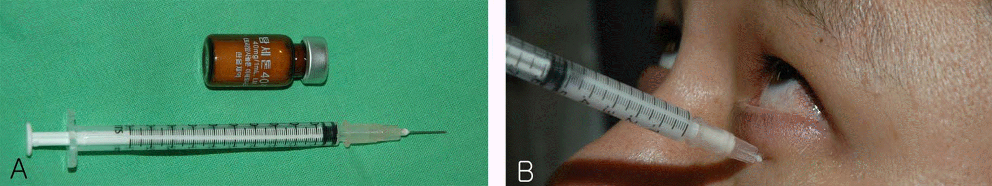 | Figure 1.Using a 26 gauge, half-inch disposable needle (A), the injection of triamcinolone acetonide of 20 mg was placed in the inferotemporal quadrant of the orbit through the lower eyelid (B). |
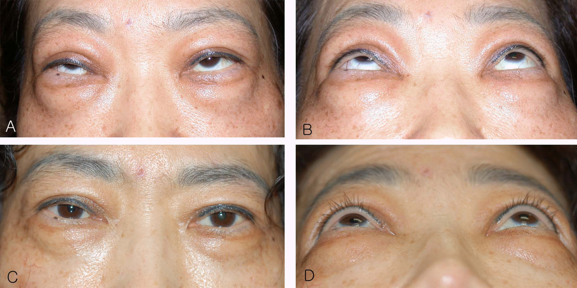 | Figure 2.Case 6. Facial photograph shows lid swelling, proptosis, hypertropia in primary gaze (A) and upgaze (B). Lid swelling, proptosis improved after periocular triamcinolone injection and ocular position was orthophoric after bilateral superior rectus recession in primary gaze (C) and upgaze (D). |
Table 1.
Summary of clinical findings
| Case No. | Age (yrs)/Sex | Duration of eye Sx∗ (mo) | Previoustherapy | Systemic steroid side effects | Thyroidstate | Thyroid therapy |
|---|---|---|---|---|---|---|
| 1 | 46/M | 3 | IV† steroids | Uncontrolled DM‡ | Hyper# | Antithyroid drugs |
| 2 | 54/F | 3 | Oral steroids | Increased body weight | Eu∗∗ | Antithyroid drugs |
| 3 | 43/M | 6 | IV steroids | Peptic ulcer, Increased blood sugar | Eu | Antithyroid drugs |
| 4 | 52/F | 12 | None | HTN§ | Eu | Antithyroid drugs |
| 5 | 43/M | 24 | Oral steroids | HTN, CHF∏ | Eu | None |
| 6 | 54/F | 36 | None | Uncountrolled DM | Hypo†† | Thyroidectomy |
Table 2.
Results of periocular triamcinolone injections
| Case No. | Improvement of soft tissue swelling | Improvement of proptosis | Improvement of motility | F/U (mo) | Additional therapy |
|---|---|---|---|---|---|
| 1 | Poor | Worsening | Poor | 7 | Radiation therapy |
| 2 | Good | No change | Poor | 10 | None |
| 3 | Poor | No change | Poor | 10 | Radiation therapy |
| 4 | Good | No change | Poor | 2 | None |
| 5 | Poor | Worsening | Poor | 14 | IV∗ steroids |
| 6 | Good | Good | Poor | 11 | Strabismus surgery |
Table 3.
The changes of extraocular muscles in 4 patients
| Before injection (mean±SD) (mm) | After injection (mean±SD) (mm) | |
|---|---|---|
| Superior muscle group∗ | 5.70±1.61 | 6.83±1.77 |
| Inferior rectus | 5.85±1.21 | 5.88±1.33 |
| Medial rectus | 6.56±0.96 | 6.60±1.33 |
| Lateral rectus | 3.94±0.93 | 4.29±1.03 |
 XML Download