

 PDF
PDF ePub
ePub Citation
 Print
Print


Abstract
Purpose
To find applicable parameters of the frequency doubling technology perimeter (FDP), which is known to be useful in detecting early visual field loss.
Methods
The subjects were 65 eyes of normal individuals, 58 eyes of glaucoma suspects, and 37 eyes of glaucoma patients. FDP (MD, PSD), SAP (MD, PSD, CPSD), and 5 parameters of OCT were analyzed. The receiver operating characteristic (ROC) curve, sensitivity, and specificity of each parameter were evaluated, and the comparison of FDP parameters by Pearson's chi-square was made between the glaucoma suspect group and the normal group. The same comparison was made among the normal+glaucoma suspect and glaucoma group.
Results
In discriminating between the normal and glaucoma suspect groups, the FDP-PSD revealed an AUC of 0.745 (cutoff: 3.17dB), which was significantly larger than that of SAP-PSD with an AUC of 0.660 (cutoff: 1.73dB). In comparing FDP, PSD by Anderson criteria was most reliable with a hit ratio of 0.732. On the other hand, in discriminating among the normal+glaucoma suspect group and glaucoma group, the SAP-PSD showed an AUC 0.971 (cutoff: 2.92dB), much larger than the FDP-PSD with an AUC of 0.863 (cutoff: 3.78dB). In addition, in comparing the parameters of FDP by Pearson chi-square, PSD 1% was the most reliable with a hit ratio of 0.813.
Go to : 
References
1. Rand A, Karim D, Sharon F, et al. Shield's Textbook of Glaucoma. 5th ed.Baltimore: Willians & Wilkins;2005. p. 1–2.
2. Medeiros FA, Sample PA, Weinreb RN. Frequency doubling technology perimetry abnormalities as predictors of glaucomatous visual field loss. Am J Ophthalmol. 2004; 137:863–71.

3. Bayer AU, Erb C. Short wavelength automated perimetry, frequency doubling technology perimetry and pattern electroretinography for prediction of progressive glaucomatous standard visual field defect. Ophthalmology. 2002; 109:1009–17.
4. Brusini P, Salvetat ML, Zeppieri M, Parisi L. Frequency doubling technology perimetry with the Humphrey matrix 30-2 test. J Glaucoma. 2006; 15:77–83.
5. Kang KD, Park CK. Comparison of diagnostic precision between preprogramed indicator and newly calculated indicator in optical coherence tomography. J Korean Ophthalmol Soc. 2006; 47:243–52.
6. Sim JO, Park CK. Optic nerve head analysis obtained by optical coherence tomography for the diagnosis of glaucoma in Koreans. J Korean Ophthalmol Soc. 2004; 45:1885–93.
7. Cello KE, Nelson-Quigg JM, Johnson CA. Frequency doubling technology perimetry for detection of glaucomatous visual field loss. Am J Ophthalmol. 2000; 129:314–22.
8. Glovinsky Y, Quigley HA, Dunkelberger GR. Retinal ganglion cell loss is size dependent in experimental glaucoma. Invest Ophthalmol Vis Sci. 1991; 32:484–91.
9. Kaplan E, Sharpley RM. The primate retina contains two types of ganglion cells, with high and low contrast sensitivity. Proc Natl Acad Sci U S A. 1986; 83:2755–7.
10. Silverman SE, Trick GI, Hart WM. Motion perception is abnormal in primary open-angle glaucoma and ocular hypertension. Invest Ophthalmol Vis Sci. 1990; 31:722–9.
11. Kelly DH. Nonlinear visual responses to flickering sinusoidal gratings. J Opt Soc Am. 1981; 71:1051–5.
12. Anderson AJ, Johnson CA. Mechanisms isolated by frequency doubling technology perimetry. Invest Ophthalmol Vis Sci. 2002; 43:398–401.
13. Morgan JE. Selective cell death in glaucoma : does it really occur? Br J Ophthalmol. 1994; 78:875–9.
14. Anderson AJ, Johnson CA. Frequency-doubling technology perimetry. Ophthalmol Clin North Am. 2003; 16:213–25.
15. Khong JJ, Dimitrov PM. Can the specificity of the FDT for glaucoma be improved by confirming abnormal results? J Glaucoma. 2001; 10:199–202.
16. Ra H, Park CK. Glaucoma discrimination by combined use of frequency doubling technology and Heidelberg retina tomography II. J Korean Ophthalmol Soc. 2005; 46:306–15.
17. Wolfs RC, Borger PH, Ramrattan RS, et al. Changing views on open-angle glaucoma: definitions and prevalences. Invest Ophthalmol Vis Sci. 2000; 41:3309–21.
18. Mastropasqua L, Brusini P, Carpineto P, et al. Humphrey matrix frequency doubling technology perimetry and optical coherence tomography measurement of the retinal nerve fiber layer thickness in both normal and ocular hypertensive subjects. J Glaucoma. 2006; 15:328–35.
19. Brusini P. Frequency doubling technology staging system 2. J Glaucoma. 2006; 15:315–20.
20. Chauhan BC, Johnson CA. Test-retest variability of frequency doubling perimetry and conventional perimetry in glaucoma patients and normal subjects. Invest Ophthalmol Vis Sci. 1999; 40:648–56.
21. Johnson CA, Samuels SJ. Screening for glaucomatous visual field loss with frequency-doubling perimetry. Invest Ophthalmol Vis Sci. 1997; 38:413–25.
22. Trible JR, Schultz RO, Tobinson JC, et al. Accuracy of glaucoma detection with frequency-doubling perimetry. Am J Ophthalmol. 2003; 110:1903–8.
23. Heeg GP, Jansonius NM. Influence of test reliability on the screening performance of frequency-doubling perimetry. Am J Ophthalmol. 2006; 141:585–7.
24. Heeg GP, Stouterbeer R, Jamsonius NM. Strategies for improving the diagnostic specificity of the frequency doubling perimeter. Acta Ophthalmol Scand. 2005; 83:53–6.
25. Horn FK, Wakili N, Unemann AM, et al. Testing for glaucoma with frequency-doubling perimetry in normals, ocular hypertensive, and glaucoma patients. Graefes Arch Clin Exp Ophthalmol. 2003; 240:658–65.
26. Gardiner SK, Anderson DR, Fingeret M, et al. Evaluation of decision rules for frequency-doubling technology screening tests. Optom Vis Sci. 2006; 83:432–7.
27. Pierre-Filho Pde T, Schimiti RB, de Vasconcellos JP, Costa VP. Sensitivity and specificity of frequency-doubling technology, tendency-oriented perimetry, SITA Standard and SITA Fast perimetry in perimetrically inexperienced individuals. Acta Ophthalmol Scand. 2006; 84:345–50.
28. Paolos F, Fabio M, Luca R, Nicola O. Detecting glaucoma with frequency-doubling technology perimetry. J Glaucoma. 2005; 14:485–91.
29. Medeiros FA, Sample PA, Zangwill LM, et al. A statistical approach to the evaluation of covariate effects on the receiver operating characteristic curves of diagnostic tests in glaucoma. Invest Ophthalmol Vis Sci. 2006; 47:2520–7.
Go to :
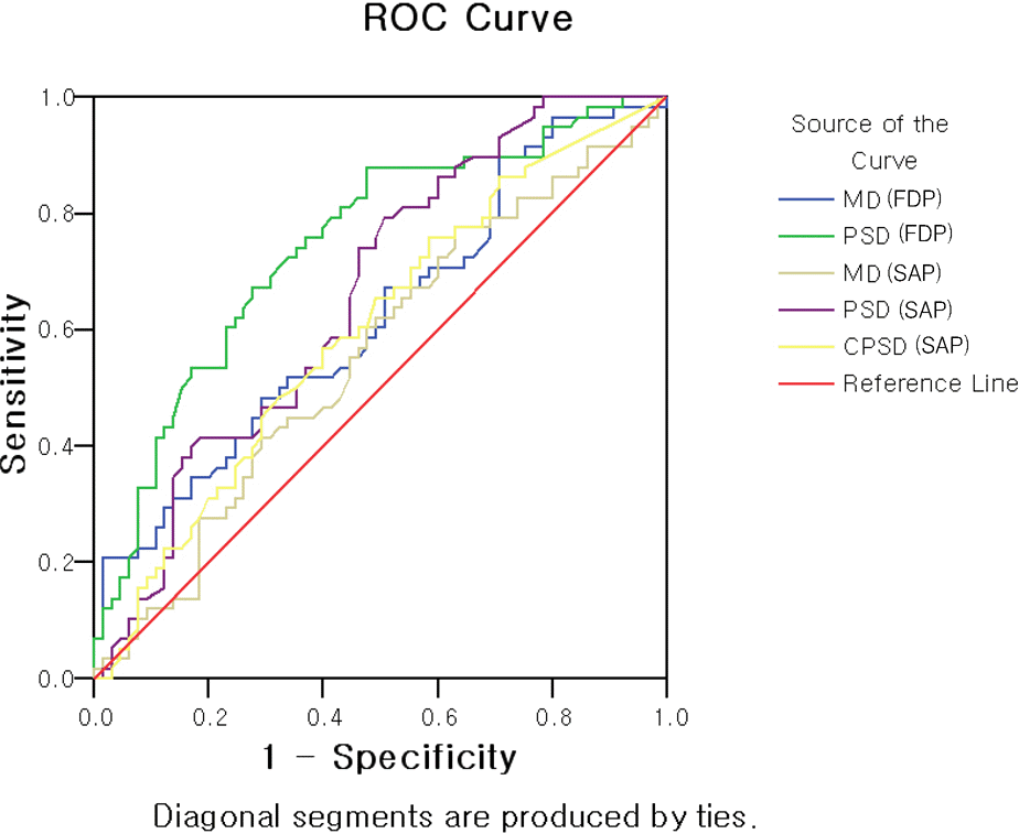 | Figure 1.ROC (receiver operator characteristic) curve of the discriminant formula. (Between glaucoma suspect and normal) |
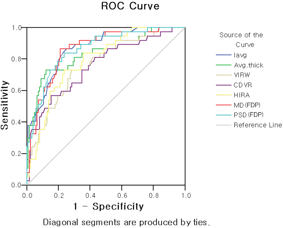 | Figure 2.ROC (receiver operator characteristic) curve of the discriminant formula. (Between ‘glaucoma’ and ‘or not’) |
Table 1.
Table 2.
| Normal | Glaucoma suspect | Glaucoma | P value | Post Hoc | |||
|---|---|---|---|---|---|---|---|
| OCT ONH | VIRWΠ | 0.27±0.21 | 0.16±0.07 | 0.11±0.07 | <0.001 | N GS | <0.001 |
| analysis | N G | <0.001 | |||||
| GS G | 0.322 | ||||||
| HIRA# | 1.59±0.27 | 1.41±0.26 | 1.22±0.23 | <0.001 | N GS | 0.001 | |
| N G | <0.001 | ||||||
| GS G | 0.002 | ||||||
| C/D | 0.62±0.10 | 0.70±0.09 | 0.76±0.11 | <0.001 | N GS | <0.001 | |
| vert.ratio | N G | <0.001 | |||||
| GS G | 0.026 | ||||||
| OCT RNFL | Iavg | 138.98±19.36 | 122.45±21.44 | 90.14±29.20 | <0.001 | N GS | 0.005 |
| analysis | N G | <0.001 | |||||
| GS G | <0.001 | ||||||
| Average | 101.13±12.11 | 93.64±12.69 | 74.71±19.07 | <0.001 | N GS | 0.016 | |
| N G | <0.001 | ||||||
| GS G | <0.001 | ||||||
| FDP | MD∗∗ | -3.74±3.73 | -6.09±5.39 | -12.43±5.61 | <0.001 | N GS | 0.030 |
| N G | <0.001 | ||||||
| GS G | <0.001 | ||||||
| PSD†† | 3.18±0.86 | 4.04±1.16 | 5.95±2.10 | <0.001 | N GS | 0.002 | |
| N G | <0.001 | ||||||
| GS G | <0.001 | ||||||
| Opthalmo | C/D | 0.47±0.15 | 0.62±0.13 | 0.70±0.14 | <0.001 | N GS | <0.001 |
| exam | hoz.ratio | N G | <0.001 | ||||
| GS G | 0.104 | ||||||
| C/D | 0.47±0.14 | 0.62±0.11 | 0.72±0.13 | <0.001 | N GS | <0.001 | |
| vert.ratio | N G | <0.001 | |||||
| GS G | 0.001 |
Table 3.
Area under the receiver operating characteristic curve (AUC), sensitivity, specificity by each parameters between glaucoma suspect and normal group
| AUC | Sensitivity (%) | Specificity (%) | Cutoff value | ||
|---|---|---|---|---|---|
| OCT∗ ONH† analysis | VIRW‡ | 0.727 | 75.90 | 63.10 | 0.205 |
| HIRA§ | 0.675 | 67.20 | 52.30 | 1.545 | |
| CDVRΠ | 0.714 | 74.10 | 63.10 | 0.648 | |
| OCT RNFL# analysis | Iavg | 0.700 | 67.20 | 40.00 | 134.500 |
| Avg thick | 0.680 | 67.20 | 40.00 | 99.320 | |
| FDP∗∗ | MD†† | 0.620 | 67.20 | 49.20 | -3.450 |
| PSD‡‡ | 0.745 | 75.90 | 63.10 | 3.170 | |
| SAP§§ | MD | 0.556 | 62.10 | 50.80 | -2.160 |
| PSD | 0.660 | 74.10 | 53.80 | 1.730 | |
| CPSDΠΠ | 0.600 | 56.90 | 60.00 | 1.190 |
Table 4.
Area under the receiver operating characteristic curve (AUC), sensitivity, specificity by each parameters between ‘glaucoma’ and ‘or not’
| AUC | Sensitivity (%) | Specificity (%) | Cutoff value | ||
|---|---|---|---|---|---|
| OCT∗ ONH† analysis | VIRW‡ | 0.761 | 71.40 | 71.50 | 0.139 |
| HIRA§ | 0.790 | 71.40 | 76.40 | 1.335 | |
| CDVRΠ | 0.765 | 77.10 | 65.00 | 0.699 | |
| OCT RNFL# analysis | Iavg | 0.876 | 88.60 | 74.80 | 118.500 |
| Avg thick | 0.832 | 85.70 | 58.50 | 95.780 | |
| FDP∗∗ | MD†† | 0.855 | 91.40 | 66.70 | -5.540 |
| PSD‡‡ | 0.863 | 88.60 | 65.90 | 3.775 | |
| SAP§§ | MD | 0.849 | 74.30 | 83.70 | -3.665 |
| PSD | 0.971 | 91.40 | 92.70 | 2.920 | |
| CPSDΠΠ | 0.959 | 94.30 | 90.20 | 1.940 |
Table 5.
Comparisons of FDP∗ by Pearson chi-square between glaucoma suspect and normal
| Hit ratio | P value | sensitivity (%) | specificity (%) | False positive | False negative | chi-square | |
|---|---|---|---|---|---|---|---|
| GHT† | 0.608 | <0.001 | 63.64 | 58.46 | 43.55 | 34.48 | 5.826 |
| MD‡ <1% | 0.585 | <0.001 | 29.31 | 84.62 | 37.04 | 42.71 | 3.469 |
| MD<5% | 0.610 | <0.001 | 53.45 | 67.69 | 40.38 | 38.03 | 5.613 |
| MD by adsΠ | 0.634 | <0.001 | 70.69 | 56.92 | 40.58 | 31.48 | 9.489 |
| PSD§<1% | 0.626 | <0.001 | 29.31 | 92.31 | 22.73 | 40.59 | 9.753 |
| PSD<5% | 0.683 | <0.001 | 62.07 | 73.85 | 32.08 | 31.43 | 16.122 |
| PSD by adsΠ | 0.732 | <0.001 | 70.69 | 75.38 | 28.07 | 25.76 | 26.166 |
| PSD by cutoff (3.17) | 0.691 | <0.001 | 75.86 | 63.08 | 35.29 | 25.45 | 18.799 |
Table 6.
Comparison of FDP∗ by Pearson chi-square between ‘glaucoma’ and ‘or not’
| Hit ratio | P value | Sensitivity π%) | Specificity π%) | False positive | False negative | chi-square | |
|---|---|---|---|---|---|---|---|
| GHT† | 0.592 | <0.001 | 94.59 | 48.33 | 63.92 | 3.33 | 22.072 |
| MD‡ <1% | 0.781 | <0.001 | 78.38 | 78.05 | 48.21 | 7.69 | 39.809 |
| MD<5% | 0.656 | <0.001 | 91.89 | 57.72 | 60.47 | 4.05 | 28.166 |
| MD by adsΠ | 0.563 | <0.001 | 97.30 | 43.70 | 65.71 | 1.82 | 21.402 |
| PSD§<1% | 0.813 | <0.001 | 78.38 | 82.11 | 43.14 | 7.34 | 47.932 |
| PSD<5% | 0.650 | <0.001 | 91.89 | 56.91 | 60.92 | 4.11 | 27.307 |
| PSD by adsΠ | 0.625 | <0.001 | 91.89 | 53.66 | 62.64 | 4.35 | 24.061 |
| PSD by cutoff (3.78) | 0.713 | <0.001 | 89.19 | 65.85 | 56.00 | 4.71 | 34.606 |
 XML Download