

 PDF
PDF ePub
ePub Citation
 Print
Print


Abstract
Purpose
To find difference in the rate of visual field defect progression among primary open angle glaucoma (POAG) patients grouped according to central corneal thickness (CCT).
Methods
The medical records of 87 eyes of 87 POAG patients who received were on regular treatment and had a minimum of 5 years of longitudinal Humphrey automated visual field follow-up were reviewed retrospectively. The patients were divided into 4 quartile groups by CCT, and the correlations among clinical factors, such as intraocular pressure (IOP), and change in mean deviation (MD), were analyzed.
Results
The thinner cornea group showed a significant trend foward higher average, maximal, and yearly maximal IOP when the IOP was corrected by CCT (p<0.01), The rate of change in MD is significantly different; −0.33±0.6dB, −0.28±0.4dB, −0.09±0.2dB,-0.02±0.2dB starting with the thinnest cornea group (p=0.036). Correlation analysis revealed a significant correlation between CCT and the rate of change in MD (r=0.351, p=0.001).
Go to : 
References
1. Damji KF, Muni RH, Munger RM. Influence of corneal variables on accuracy of intraocular pressure measurements. J Glaucoma. 2003; 12:69–80.
2. Shah S, Chatterjee A, Mathai M, et al. Relationship between corneal thickness and measured intraocular pressure in a general ophthalmology clinic. Ophthalmology. 1999; 106:2154–60.
3. Ehlers N, Bramsen T, Sperling S. Applanation tonometer and central corneal thickness. Acta Ophthalmol. 1967; 77:734–40.
4. Whitacre MM, Stein RA, Hassanein K. The effect of corneal thickness on applanation tonometry. Am J Ophthalmol. 1993; 115:592–6.

5. Copt RP, Thomas R, Mermound A. Corneal thickness in ocular hypertension, primary open-angle glaucoma, and normal tension glaucoma. Arch Ophthalmol. 1999; 117:14–6.
6. Gordon MO, Beiser JA, Brandt JD, et al. The Ocular Hypertension Treatment Study: baseline factors that predict the onset of primary open-angle glaucoma. Arch Ophthalmol. 2002; 120:714–20.
7. Brandt JD, Beiser JA, Kass MA, et al. Central corneal thickness in the Ocular Hypertension Treatment Study (OHTS). Ophthalmology. 2001; 108:1779–88.
8. Kass MA, Hart WH Jr, Gorgon M, Miller JP. Risk factors favoring the development of glaucomatous visual field loss in ocular hypertension. Surv Ophthalmol. 1980; 25:155.
9. Herndon LW, Weizer JS, Stinnett SS. Central corneal thickness as a risk factor for advanced glaucoma damage. Arch Ophthalmol. 2004; 122:17–21.
10. Doughty MJ, Zaman ML. Human corneal thickness and its impact on intraocular pressure measures: a review and meta-analysis approach. Surv Ophthalmol. 2000; 44:367–408.
11. Congdon NG, Broman AT, Bandeen-Rochek , et al. Central corneal thickness and corneal hysteresis associated with glaucoma damage. Am J Ophthalmol. 2006; 141:868–75.
12. Hansen FK. A clinical study of the normal human central corneal thickness. Acta Ophthalmol. 1971; 49:82–9.
13. Shah S, Spedding C, Bhojwani R, et al. Assessment of the diurnal variation in central corneal thickness and intraocular pressure for patients with suspected glaucoma. Ophthalmology. 2000; 107:1191–3.
14. Argus WA. Ocular hypertension and central corneal thickness. Ophthalmology. 1995; 102:1810–2.
15. Medeiros FA, Sample PA, Zangwill LM, et al. Corneal thickness as a risk factor for visual field loss in patients with preperimetric glaucomatous optic neuropathy. Am J Ophthalmol. 2003; 136:805–13.
16. Kim JW, Chen PP. Central corneal pachymetry and visual field progress in patients with open-angle glaucoma. Ophthalmology. 2004; 111:2126–32.
17. Choi YJ, Kim JH, Sohn YH. Influence of central corneal thickness on diagnosis of glaucoma. J Korean Ophthalmol Soc. 2003; 44:2823–8.
Go to :
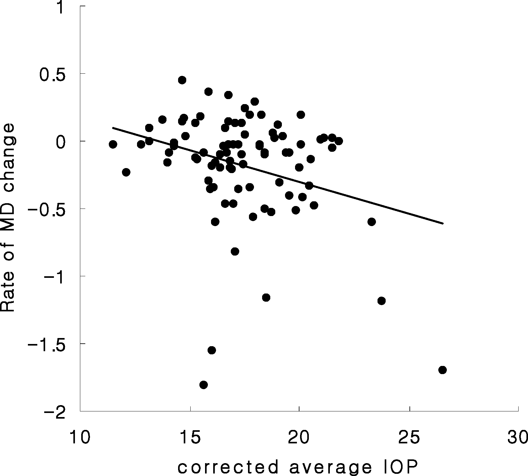 | Figure 1.Scattergram of changes in mean deviation in automated visual field test and corrected intraocular pressure with central corneal thickness (Pearson correlation test, r=-0.302, p=0.004). Rate of MD change (dB/yr)=(final MD - initial MD) / follow-up years. |
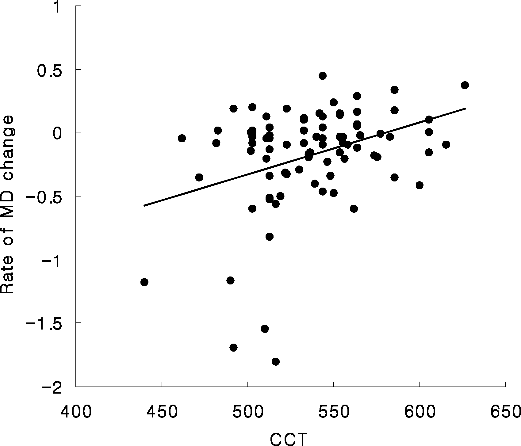 | Figure 2.Scattergram of central corneal thickness and change in mean deviation in automated visual field test (Pearson correlation test, r=0.351, p=0.001). Rate of MD change (dB/yr)=(final MD - initial MD) / follow-up years. |
Table 1.
Demographics of the patients
Table 2.
Patient characteristics grouped by central corneal thickness
(mean±SD)
| Group (n) | Group 1 (20) | Group 2 (23) | Group 3 (22) | Group 4 (22) | p-value∗ |
|---|---|---|---|---|---|
| CCT | <513 μm | 513∼535 μm | 536∼557 μm | >557 μm | |
| Follow up (years) | 7.3±2.5 | 6.4±1.6 | 8.5±3.7 | 7.1±2.2 | 0.077 |
| Age (years) | 61.1±9.8 | 58.0±9.7 | 55.5±7.4 | 58.4±10.5 | 0.301 |
| Sex (male/female) | 14/6 | 15/8 | 17/5 | 13/9 | 0.630 |
| Laterality (Right/Left) | 12/8 | 8/15 | 14/8 | 14/8 | 0.153 |
| Systemic diseases (No) | 0.4±0.5 | 0.3±0.5 | 0.7±0.8 | 0.5±0.7 | 0.179 |
| Number of surgery | 0.8±1.2 | 0.7±0.8 | 0.5±0.9 | 0.6±0.9 | 0.857 |
Table 3.
Comparison of intraocular pressure course and changes of cup-disc ratio in 4 groups divided by central corneal thickness
(mean±SD)
| Group (n) | Group 1 (20) | Group 2 (23) | Group 3 (22) | Group 4 (22) | p-value∗ |
|---|---|---|---|---|---|
| CCT | <513 μm | 513∼535 μm | 536∼557 μm | >557 μm | |
| IOP on 1st visit | 21.2±3.8 | 22.2±5.5 | 19.9±4.0 | 21.5±4.6 | 0.397 |
| Glaucoma meds. | 1.5±1.2 | 1.7±0.9 | 1.7±1.0 | 1.3±1.2 | 0.688 |
| on 1st visit | |||||
| Average IOP | 17.4±1.7 | 17.1±2.4 | 16.9±1.9 | 18.5±2.2 | 0.052 |
| Highest IOP | 23.0±4.6 | 25.8±6.0 | 22.7±4.8 | 23.9±4.7 | 0.186 |
| Yearly max IOP | 19.2±3.6 | 19.8±2.6 | 18.3±2.3 | 19.9±2.5 | 0.210 |
| Yearly variation | 4.0±3.1 | 4.8±2.4 | 3.1±1.4 | 3.6±1.4 | 0.060 |
| Average No. of | 1.8±0.8 | 1.8±0.6 | 1.5±0.9 | 1.4±0.8 | 0.268 |
| glaucoma meds. | |||||
| Initial C/D ratio | 0.78±0.2 | 0.80±0.2 | 0.77±0.1 | 0.69±0.2 | 0.121 |
| Last C/D ratio | 0.87±0.1 | 0.88±0.1 | 0.87±0.1 | 0.78±0.2 | 0.065 |
| Change of C/D ratio | 0.08±0.1 | 0.08±0.1 | 0.10±0.1 | 0.09±0.1 | 0.894 |
Table 4.
Comparison of corrected intraocular pressure according to the central corneal thickness in 4 groups divided by central corneal thickness
(mean± SD)
| Group (n) | Group 1 (20) | Group 2 (23) | Group 3 (22) | Group 4 (22) | p-value∗ |
|---|---|---|---|---|---|
| CCT | <513 μm | 513∼535 μm | 536∼557 μm | <557 μm | |
| Average IOP | 19.5±3.0 | 18.2±1.6 | 16.3±2.0 | 15.9±2.2 | 0.000 |
| Highest IOP | 26.5±6.1 | 25.1±4.8 | 22.2±4.9 | 21.6±4.8 | 0.005 |
| Yearly maximal IOP | 21.4±3.8 | 20.6±2.6 | 17.8±2.3 | 17.7±2.5 | 0.000 |
Table 5.
Comparison of change in mean deviation in Humphrey automated visual field in 4 groups divided by central corneal thickness
(mean± SD)
| Group (n) | Group 1 (20) | Group 2 (23) | Group 3 (22) | Group 4 (22) | p-value∗ |
|---|---|---|---|---|---|
| CCT | <513 μm | 513∼535 μm | 536∼557 μm | >557 μm | |
| MD Initial | -12.4±10.1 | -11.0±8.5 | -10.0±8.3 | -6.0±7.1 | 0.089 |
| MD Last | -14.2±11.3 | -12.9±9.3 | -10.6±8.0 | -6.1±7.2 | 0.022 |
| MD Change | -1.9±3.4 | -1.9±3.2 | -0.6±1.9 | -0.2±1.3 | 0.056 |
| MD Slope† | -0.33±0.6 | -0.28±0.4 | -0.09±0.2 | -0.02±0.2 | 0.036 |
 XML Download