

 PDF
PDF ePub
ePub Citation
 Print
Print


Abstract
Purpose
To determine the factors influence retinal nerve fiber layer (RNFL) and optic nerve head (ONH) parameters measured by Stratus optical coherence tomography (OCT).
Methods
Topographic RNFL thickness and optic disc parameters of 129 healthy Korean subjects of aged 14 to 87 were measured using the fast retinal nerve fiber layer thickness and fast optic disk algorithms of Stratus OCT. One eye of each subject was randomly selected for statistical analysis. Using multiple linear regression, the effect of optic disc area, age, refractive error, and zone beta on each parameter was analyzed.
Results
Large discs had large horizontal integrated rim width (HIRW), cup area, rim area, C/D area ratio, and vertical C/D ratio. The thickness of average, superior, inferior, and nasal quadrant RNFL increased significantly with an increase in optic disc area. Average and superior quadrant RNFL thickness, and HIRW decreased with age. Refractive error showed a correlation with the vertical integrated rim area, horizontal C/D ratio, and temporal quadrant RNFL thickness. Gender and zone beta had no statistically significant influence on ONH and RNFL parameters.
Go to : 
References
1. Paunescu LA, Schuman JS, Price LL, et al. Reproducibility of nerve fiber thickness, macular thickness, and optic nerve head measurements using Stratus OCT. Invest Ophthalmol Vis Sci. 2004; 45:1716–24.
2. Bowd C, Zangwill LM, Blumenthal EZ, et al. Imaging of the optic disc and retinal nerve fiber layer: the effects of age, optic disc area, refractive error, and gender. J Opt Soc Am A Opt Image Sci Vis. 2002; 19:197–207.

3. Durukan AH, Yucel I, Akar Y, Bayraktar MZ. Assessment of optic nerve head topographic parameters with a confocal scanning laser ophthalmoscope. Clin Experiment Ophthalmol. 2004; 32:259–64.
4. Nakamura H, Maeda T, Suzuki Y, Inoue Y. Scanning laser tomography to evaluate optic discs of normal eyes. Jpn J Ophthalmol. 1999; 43:410–4.
5. Park HJ, Hwang JH, Uhm KB. The influence of age, gender, refractive error, and optic disc area on the HRT parameters in normal eyes. J Korean Ophthalmol Soc. 2004; 45:982–9.
6. Saruhan A, Org lS, Kocak I, et al. Descriptive information of topographic parameters computed at the optic nerve head with the Heidelberg Retina Tomograph. J Glaucoma. 1998; 7:420–9.
7. Savini G, Zanini M, Carelli V, et al. Correlation between retinal nerve fiber layer thickness and optic nerve head size: an optical coherence tomography study. Br J Ophthalmol. 2005; 89:489–92.
8. Hougaard JL, Ostenfeld C, Heijl A, Bengtsson B. Modelling the normal retinal nerve fibre layer thickness as measured by Stratus optical coherence tomography. Graefes Arch Clin Exp Ophthalmol. 2006; 417:372–9.
9. Varma R, Skaf M, Barron E. Retinal nerve fiber layer thickness in normal human eyes. Ophthalmology. 1996; 103:2114–9.
10. Jonas JB, Budde WM, Panda-Jonas S. Ophthalmoscopic evaluation of the optic nerve head. Surv Ophthalmol. 1999; 43:293–320.
11. Schuman JS, Pedut-Kloizman T, Hertzmark E, et al. Reproducibility of nerve fiber layer thickness measurements using optical coherence tomography. Ophthalmology. 1996; 103:1889–98.
12. Jonas JB, Schmidt AM, Muller-Bergh JA, et al. Human optic nerve fiber count and optic disc size. Invest Ophthalmol Vis Sci. 1992; 33:2012–8.
13. Ramrattan RS, Wolfs RCW, Jonas JB, et al. Determinants of optic disc characteristics in a general population: The Rotterdam Study. Ophthalmology. 1999; 106:1588–96.
14. Varma R, Tielsch JM, Quigley HA, et al. Race-, age-, gender-, and refractive error-related differences in the normal optic disc. Arch Ophthalmol. 1994; 112:1068–76.
15. Repka MX, Quigley HA. The effect of age on normal human optic nerve fiber number and diameter. Ophthalmology. 1989; 96:26–32.
16. Moya FJ, Brigatti L, Caprioli J. Effect of aging on optic nerve appearance: a longitudinal study. Br J Ophthalmol. 1999; 83:567–72.
17. Weinreb RN, Shakiba S, Zangwill L. Scanning laser polarimetry to measure the nerve fiber layer of normal and glaucomatous eyes. Am J Ophthalmol. 1995; 119:627–36.
18. Chi QM, Tomita G, Inazumi K, et al. Evaluation of the effect of aging on the retinal nerve fiber layer thickness using scanning laser polarimetry. J Glaucoma. 1995; 4:406–13.
19. Hougaard JL, Kessel L, Sander B, et al. Evaluation of heredity as a determinant of retinal nerve fiber layer thickness as measured by optical coherence tomography. Invest Ophthalmol Vis Sci. 2003; 44:3011–6.
20. Kanamori A, Escano MF, Eno A, et al. Evaluation of the effect of aging on retinal nerve fiber layer thickness measured by optical coherence tomography. Ophthalmologica. 2003; 217:273–8.
21. Mikelberg FS, Yidegiligne HM, White VA, et al. Relation between optic nerve axon number and axon diameter to scleral canal area. Ophthalmology. 1991; 98:60–3.
22. Iliev ME, Meyenberg A, Garweg JG. Morphometric assessment of normal, suspect and glaucomatous optic discs with Stratus OCT and HRT II. Eye. 2006; 20:1288–99.
23. Schuman JS, Wollstein G, Farra T, et al. Comparison of optic nerve head measurements obtained by optical coherence tomography and confocal scanning laser ophthalmoscopy. Am J Ophthalmol. 2003; 135:504–12.
24. Jonas JB, Gusek GC, Naumann GOH. Optic disc, cup and neuroretinal rim size, configuration and correlations in normal eyes. Invest Ophthalmol Vis Sci. 1988; 29:1151–8.
25. Heijl A, Molder H. Optic disc diameter influences the ability to detect glaucomatous disc damage. Acta Ophthalmol. 1993; 71:122–9.
26. Jonas JB, Gusek GC, Naumann GOH. Optic disk morphometry in high myopia. Graefes Arch Clin Exp Ophthalmol. 1988; 226:587–90.
27. Rudnicka AR, Frost C, Owen CG, Edgar DF. Nonlinear behavior of certain optic nerve head parameters and their determinants in normal subjects. Ophthalmology. 2001; 108:2358–68.
28. Hoh ST, Lim MC, Seah SK, et al. Peripapillary retinal nerve fiber layer thickness variations with myopia. Ophthalmology. 2006; 113:773–7.
29. Bayraktar S, Bayraktar Z, Yilmaz OF. Influence of scan radius correction for ocular magnification and relationship between scan radius with retinal nerve fiber layer thickness measured by optical coherence tomography. J Glaucoma. 2001; 10:163–9.
30. Yamazaki Y, Yoshikawa K, Kunimatsu S, et al. Influence of myopic disc shape on the diagnostic precision of the Heidelberg Retina Tomograph. Jpn J Ophthalmol. 1999; 43:392–7.
31. Blumenthal EZ, Williams JM, Weinreb RN, et al. Reproducibility of nerve fiber layer thickness measurements by use of optical coherence tomography. Ophthalmology. 2000; 107:2278–82.
32. Carpineto P, Ciancaglini M, Zuppardi E, et al. Reliability of nerve fiber layer thickness measurements using optical coherence tomography in normal and glaucomatous eyes. Ophthalmology. 2003; 110:190–5.
33. Gundersen KG, Heijl A, Bengtsson BO. Age, gender, IOP, refraction and optic disc topography in normal eyes. A cross-sectional study using raster and scanning laser tomography. Acta Ophthalmol Scand. 1998; 76:170–5.
34. Vernon SA, Hawker MJ, Ainsworth G, et al. Laser scanning tomography of the optic nerve head in a normal elderly population: The Bridlington eye assessment project. Invest Ophthalmol Vis Sci. 2005; 46:2823–8.
Go to :
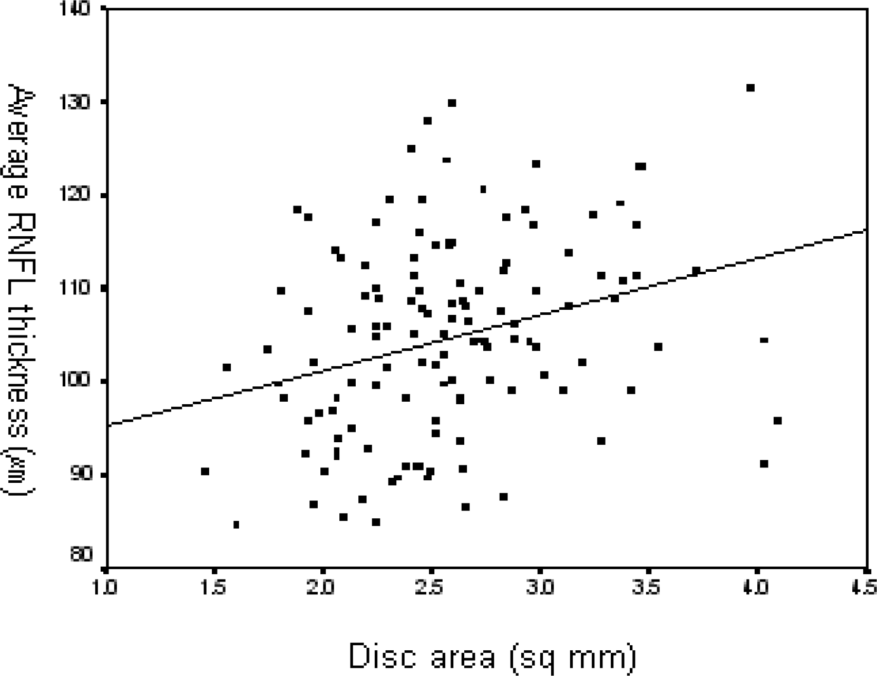 | Figure 1.Simple linear regression (y=89.5+5.954xdisc area, R=0.298, R2=8.9%, P=0.001) between disc area and average retinal nerve fiber layer (RNFL) thickness. |
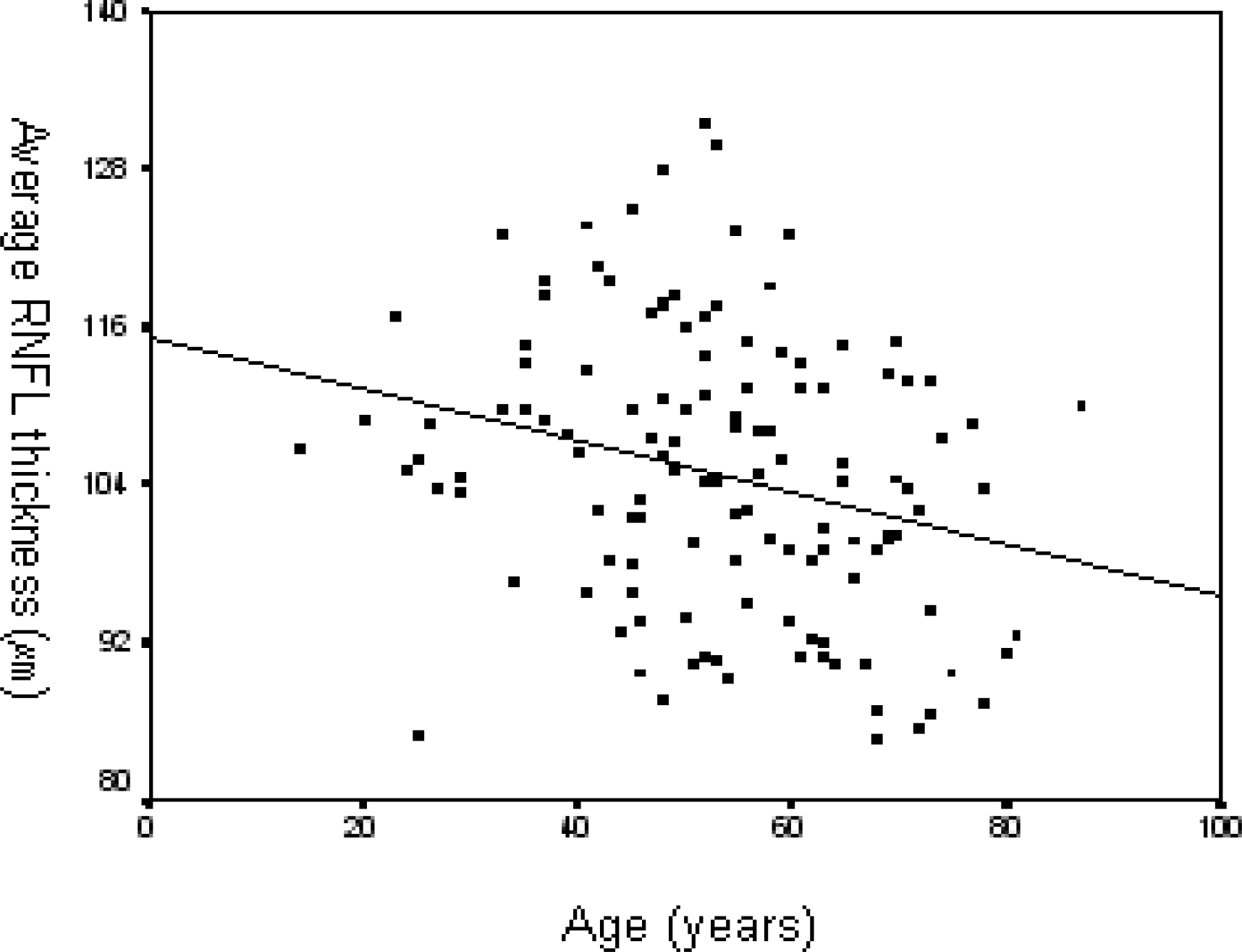 | Figure 2.Simple linear regression (y=115.3-0.197xage, R=0.268, R2=7.2%, P=0.002) between age and average retinal nerve fiber layer (RNFL) thickness. |
Table 1.
The influence of gender on optic disc and retinal nerve fiber layer parameters (mean value±standard deviation)
Table 2.
The influence of zone beta on optic disc and retinal nerve fiber layer parameters (mean value±standard deviation)
Table 3.
Effect of optic disc area, age, and refractive error on optic disc and retinal nerve fiber layer parameters analyzed with simple linear regression
Table 4.
Effect of optic disc area, age, and refraction on optic disc and retinal nerve fiber layer parameters analyzed with multiple linear regression model
Table 5.
Effect of optic disc area, age, and refraction on optic disc and retinal nerve fiber layer parameters analyzed with multiple linear regression model
 XML Download