Abstract
Purpose
To compare results between in femtosecond laser and microkeratome LASIK correction of myopia
Methods
We retrospectively analyzed the result of 94 eyes of 47 patients in the femtosecond group (F) and 103 eyes of 52 patients in the microkeratome group (M). All patients had undergone LASIK using either a femtosecond laser or a microkeratome for making of flap. Patients were divided into groups I (6D≤) and II (≥6D) according to preoperative myopia. Each patient was followed up for over 6 months with measurements of uncorrected visual acuity and manifest refraction at 1 week and 1, 3, and 6 months after operation. Complications during and after the operation were reviewed retrospectively in two groups 6month after the operation.
Results
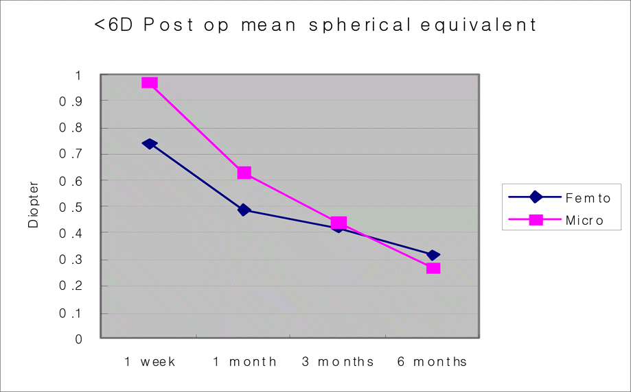
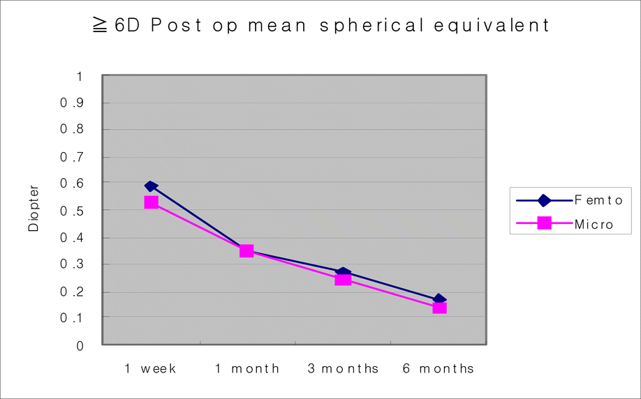
In groups F-I, F-II, M-I, and M-II, postoperative 6-month uncorrected visual acuity was 0.98±0.08, 0.96±0.09, 0.97±0.03, 0.98±0.09. At the 6-month follow-up, there were no significant differences between the two groups in uncorrected visual acuity and mean spherical equivalent. Corneal opacity was found in 3 eyes in group M and complication related with flap was found 1 eye in group F and 4 eyes in group M.
Go to : 
References
2. Ratkay-Traub I, Juhasz T, Horvath C, et al. Ultra-short pulse (femtosecond) laser surgery; initial use in LASIK flap creation. Ophthalmol Clin North Am. 2001; 14:347–55.
3. Nordan LT, Slade SG, Baker RN, et al. Femtosecond laser flap creation for laser in situ keratomileusis: six-month follow-up of initial US clinical series. J Refract Surg. 2003; 19:8–14.

4. Kurtz RM, Elner V, Liu X, et al. Plasma-mediated ablation of biological tissue with picosecond and femtosecond laser pulses. Proc SPIE. 1997; 2975:192–200.
5. Shemesh G, Dotan G, Lipshitz I. Predictability of corneal flap thickness in laser in situ keratomelusis using three different microkeratomes. J Refract Surg. 2002; 18:S347–51.

6. Flanagan GF, Binger PS. Precision of flap measurements for laser in situ keratomileusis in 4428 eyes. J Refract Surg. 2003; 19:113–23.

7. Juhasz T, Loesel FH, Kurtz RM, et al. Corneal refractive surgery with femtosecond lasers. IEEE J Select Topics Quantum Electron. 1999; 5:902–10.

8. Tham VMB, Maloney RK. Microkeratome complications of laser in situ keratomileusis. Ophthalmology. 2000; 107:920–4.

9. Walker MB, Wilson SE. Lower intraoperative flap complication rate with the Hansatome microkeratome compared to the Automate corneal shaper. J Refract Surg. 2000; 16:79–82.
10. Yoon JT, Lee GJ, Tchah HW. Flap complication of LASIK. J Korean Ophthalmol Soc. 2000; 41:1146–50.
11. Jacobs JM, Travella MJ. Incidence of intraoperative flap complications in laser in situ keratomileusis. J Cataract Refract Surg. 2002; 28:23–8.

12. Hanna KD, poulique YM, Waring GO III, et al. Corneal wound healing in monkeys after repeated excimer laser photorefractive keratectomy. Arch Ophthalmol. 1992; 110:1286–91.

13. Karl GS, Jon GD, Ignacio TS, Binder PS. Transient light sensitivity after femtosecond laser flap creation: Clinical findings and management. J Cataract Refract Surg. 2006; 32:91–4.
14. Juhasz T, Kastis GA, Suarez C, et al. Time-resolved observations of shock waves and cavitation bubbles generated by femtosecond laser pulses in corneal tissue and water. Lasers Surg Med. 1996; 19:23–31.

15. Juhasz T, Djotyan G, Loesel FH, et al. Application of femtosecond lasers in corneal surgery. Laser Phys. 2000; 10:495–500.
16. Loesel FH, Niemz MH, Bille JF, Juhasz T. Laser-induced optical breakdown on hard and soft tissues and its dependence on the pulse duration:experiment and model. IEEE J Quantum Electron. 1996; 32:1717–22.
17. Binder PS. Flap dimensions created with the IntraLase FS laser. J Cataract Refract Surg. 2004; 30:26–32.

18. Daniel SD, Guy MK. Femtosecond laser versus mechanical keratome flaps in wave front-guided laser in situ keratomileusis. J Cataract Refract Surg. 2005; 31:120–6.
19. Guy MK, Karl GS. Comparison of the IntraLase femtosecond laser and mechanical keratomes for laser in situ keratomileusis. J Cataract Refract Surg. 2004; 30:804–11.
Go to : 
 | Figure 1.Postoperative mean spherical equivalent of <6D Femtosecond and Microkeratome groups. |
 | Figure 2.Postoperative mean spherical equivalent of ≧6D Femtosecond and Microkeratome groups. |
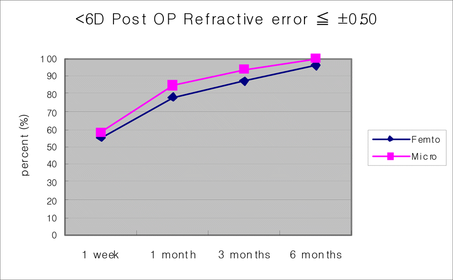
 | Figure 3.Postoperative Refractive error ≦±0.50 of <6D Femtosecond and Microkeratome groups. |
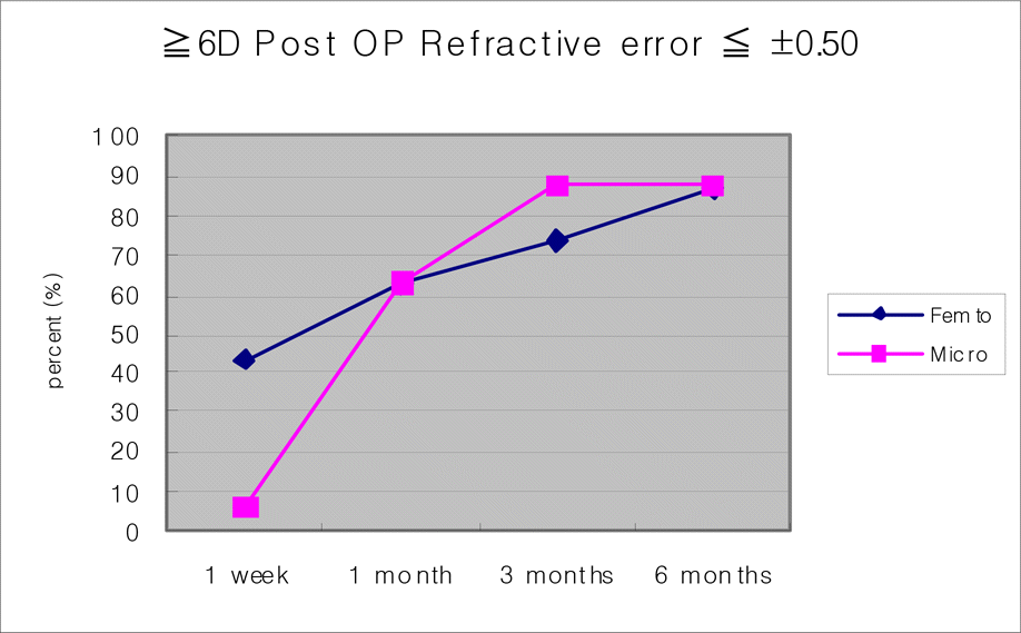
 | Figure 4.Postoperative refractive error ≦±0.50 of ≧6D Femtosecond and Microkeratome groups. |
Table 1.
Summary of data about patient who had LASIK operation using Femtosecond laser and Microkeratome
| Femtosecond | Microkeratome | |
|---|---|---|
| Age (years) | 26.53±5.11 | 25.16±2.89 |
| Male: Female (eyes) | 33:67 | 31:69 |
| Pre-op. IOP†(mmHg) | 13.43±2.34 mmHg | 12.75±2.11 mmHg |
| Pre-op SE∗ (D) | −5.90±1.76 (−3.25∼−8.75) | −4.26±1.74D (−2.52∼−6.00) |
| Pre-op K (mm) | 44.51±1.32 (41.10∼47.54) | 43.54±1.44 (40.68∼46.58) |
| Corneal thickness (µm) | 507.41±31.55 | 523.22±31.55 |
| Total patients (eyes) | 52 (100) | 55 (100) |
Table 2.
Uncorrected visual acuity (UCVA) after Femtosecond laser and Microkeratome
Table 3.
Refractive errors after Femtosecond laser and Microkeratome


 PDF
PDF ePub
ePub Citation
 Print
Print


 XML Download