

 PDF
PDF ePub
ePub Citation
 Print
Print


Abstract
We report a case of acute pancreatitis secondary to pancreatic neuroendocrine tumor. A 46-year old man presented with upper abdominal pain. The serum amylase and lipase were elevated. Abdominal computed tomography (CT) and magnetic resonance cholangiopancreatography revealed a 1.7 cm sized mass at the pancreas body with a dilatation of the upstream pancreatic duct and mild infiltrations of peripancreatic fat. An endoscopic ultrasound-guided fine needle biopsy was performed for the pancreatic mass, but only necrotic tissue was observed on the pathologic examination. A chest and neck CT scan revealed anterior mediastinal, paratracheal, and cervical lymph node enlargement, which were indicative of metastasis. An ultrasound-guided core needle biopsy was performed for the enlarged neck lymph node, and pathologic examination revealed a metastatic poorly differentiated carcinoma. Immunohistochemical analysis showed positive staining for synaptophysin, chromogranin A, and CD 56, indicative of a neuroendocrine carcinoma.
Figures and Tables
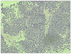
Fig. 1
(A) Abdominal computed tomography showed a 1.7 cm sized low attenuated lesion at the pancreatic body (white arrow), peripancreatic infiltration (arrowheads), adrenal nodule (black arrow), and (B) p-duct dilation (black arrowhead).
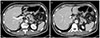
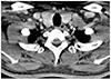
Fig. 2
Magnetic resonance cholangiopancreatography showed dilation of the upstream pancreatic duct (white arrow).
References
1. Jin SY. Recent update of pathology of the pancreatic neuroendocrine tumor. Korean J Med. 2011; 80:371–377.
2. Chang HM. Consensus guideline for advanced pancreatic neuroendocrine tumor. Korean J Med. 2011; 80:393–396.
3. Kimura W, Kuroda A, Morioka Y. Clinical pathology of endocrine tumors of the pancreas. Analysis of autopsy cases. Dig Dis Sci. 1991; 36:933–942.
4. Viúdez A, De Jesus-Acosta A, Carvalho FL, Vera R, Martín-Algarra S, Ramírez N. Pancreatic neuroendocrine tumors: challenges in an underestimated disease. Crit Rev Oncol Hematol. 2016; 101:193–206.

5. Tejedor Bravo M, Justo LM, Lasala JP, Moreira Vicente VF, Ruiz AC, Scapa Mde L. Acute pancreatitis secondary to neuroendocrine pancreatic tumors: report of 3 cases and literature review. Pancreas. 2012; 41:485–489.
6. Ahn SS, Kim MJ, Kim D. An insidious pancreatic lesion in a young woman with recurrent pancreatitis. Gastroenterology. 2010; 139:e9–e10.
7. Maimone S, Agrawal D, Pollack MJ, et al. Variability in measurements of pancreatic cyst size among EUS, CT, and magnetic resonance imaging modalities. Gastrointest Endosc. 2010; 71:945–950.
8. Griñó P, Martínez J, Griñó E, et al. Acute pancreatitis secondary to pancreatic neuroendocrine tumours. JOP. 2003; 4:104–110.
9. Jang DK, Park JM, Lee M, et al. A Case of Long-term Survivor of Pancreatic Neuroendocrine Tumor with Multiple Liver Metastases. Korean J Pancreas Biliary Tract. 2013; 18:62–66.
10. Tsutsumi K, Ohtsuka T, Mori Y, et al. Analysis of lymph node metastasis in pancreatic neuroendocrine tumors (PNETs) based on the tumor size and hormonal production. J Gastroenterol. 2012; 47:678–685.
11. Kawamoto S, Shi C, Hruban RH, et al. Small serotonin-producing neuroendocrine tumor of the pancreas associated with pancreatic duct obstruction. AJR Am J Roentgenol. 2011; 197:W482–W488.
12. Hamada Y, Nakayama Y, Maeshiro K, Ikeda T, Hayashi H, Iwasaki H. Two cases of primary carcinoid tumor of the pancreas associated with marked stenosis of the main pancreatic duct. Pancreas. 2009; 38:834–835.
13. Dhir V, Joshi N, Vivekanandarajah S, Bhandari S, Bapat M, Maydeo A. Recurrent acute pancreatitis in a patient with wirsungocele and neuroendocrine tumor of ampulla of Vater. JOP. 2013; 14:99–101.
14. Oh TG, Bang S. Pathogenesis of acute pancreatitis. Korean J Med. 2013; 85:111–115.
15. Pavel ME, Hassler G, Baum U, Hahn EG, Lohmann T, Schuppan D. Circulating levels of angiogenic cytokines can predict tumour progression and prognosis in neuroendocrine carcinomas. Clin Endocrinol (Oxf). 2005; 62:434–443.
16. Blogowski W, Deskur A, Budkowska M, et al. Selected cytokines in patients with pancreatic cancer: a preliminary report. PLoS One. 2014; 9:e97613.
 XML Download