

 PDF
PDF ePub
ePub Citation
 Print
Print


References
1. Cronkhite LW Jr, Canada WJ. Generalized gastrointestinal polyposis; an unusual syndrome of polyposis, pigmentation, alopecia and onychotrophia. New Engl J Med. 1955; 252:1011–1015.
2. Watanabe C, Komoto S, Tomita K, et al. Endoscopic and clinical evaluation of treatment and prognosis of Cronkhite-Canada syndrome: a Japanese nationwide survey. J Gastroenterol. 2016; 51:327–336.

3. Wen XH, Wang L, Wang YX, Qian JM. Cronkhite-Canada syndrome: report of six cases and review of literature. World J Gastroenterol. 2014; 20:7518–7522.
4. Kang DU, Yang DH, Choi Y, et al. A case of Cronkhite-Canada syndrome showing spontaneous remission. Intest Res. 2013; 11:317–322.
5. Yun SH, Cho JW, Kim JW, et al. Cronkhite-Canada syndrome associated with serrated adenoma and malignant polyp: a case report and a literature review of 13 Cronkhite-Canada syndrome cases in Korea. Clin Endosc. 2013; 46:301–305.
6. Daniel ES, Ludwig SL, Lewin KJ, Ruprecht RM, Rajacich GM, Schwabe AD. The Cronkhite-Canada syndrome. An analysis of clinical and pathologic features and therapy in 55 patients. Medicine (Baltimore). 1982; 61:293–309.
7. Sweetser S, Ahlquist DA, Osborn NK, et al. Clinicopathologic features and treatment outcomes in Cronkhite-Canada syndrome: support for autoimmunity. Dig Dis Sci. 2012; 57:496–502.
Go to : 
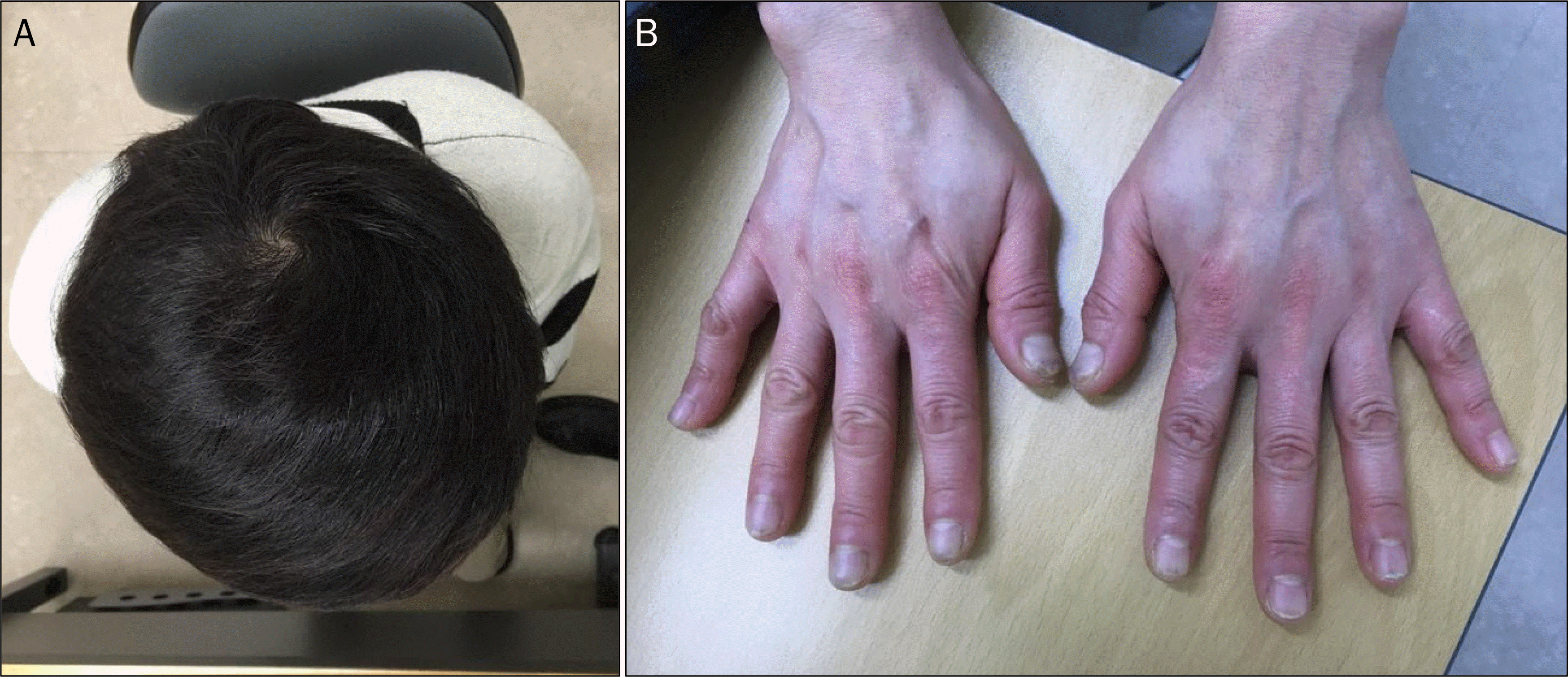
 | Fig. 1.Clinical manifestation of the patient at the initial presentation. (A) Hair loss. (B) Skin pigmentation and nail dystrophy. (C) Tongue swelling and mucosal atrophy. |
 | Fig. 2.Initial upper and lower endoscopic findings. Numerous variable-sized sessile polyps with mucosal edema and hyperemia were observed in the (A, B) stomach, and (C, D) colon. |
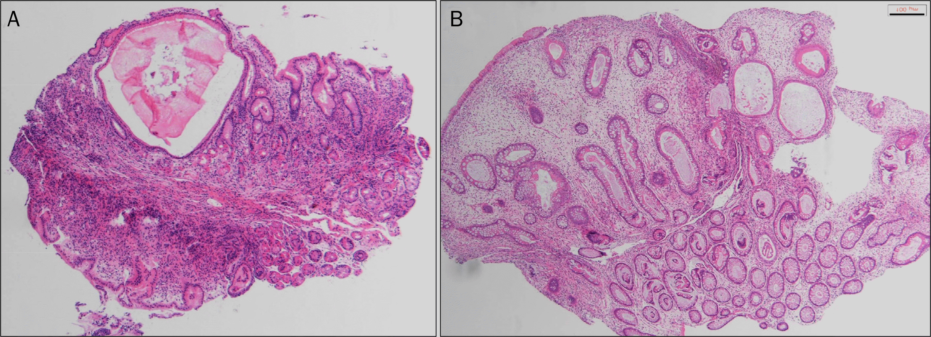 | Fig. 3.Histologic findings. (A) Dilatation of gland, and inflammatory cell infiltration in gastric mucosa (H&E, ×40). (B) Dilatation of gland, cystic dilatation of crypt, and edema of lamina propria in colonic mucosa (H&E, ×40). |

 XML Download