Abstract
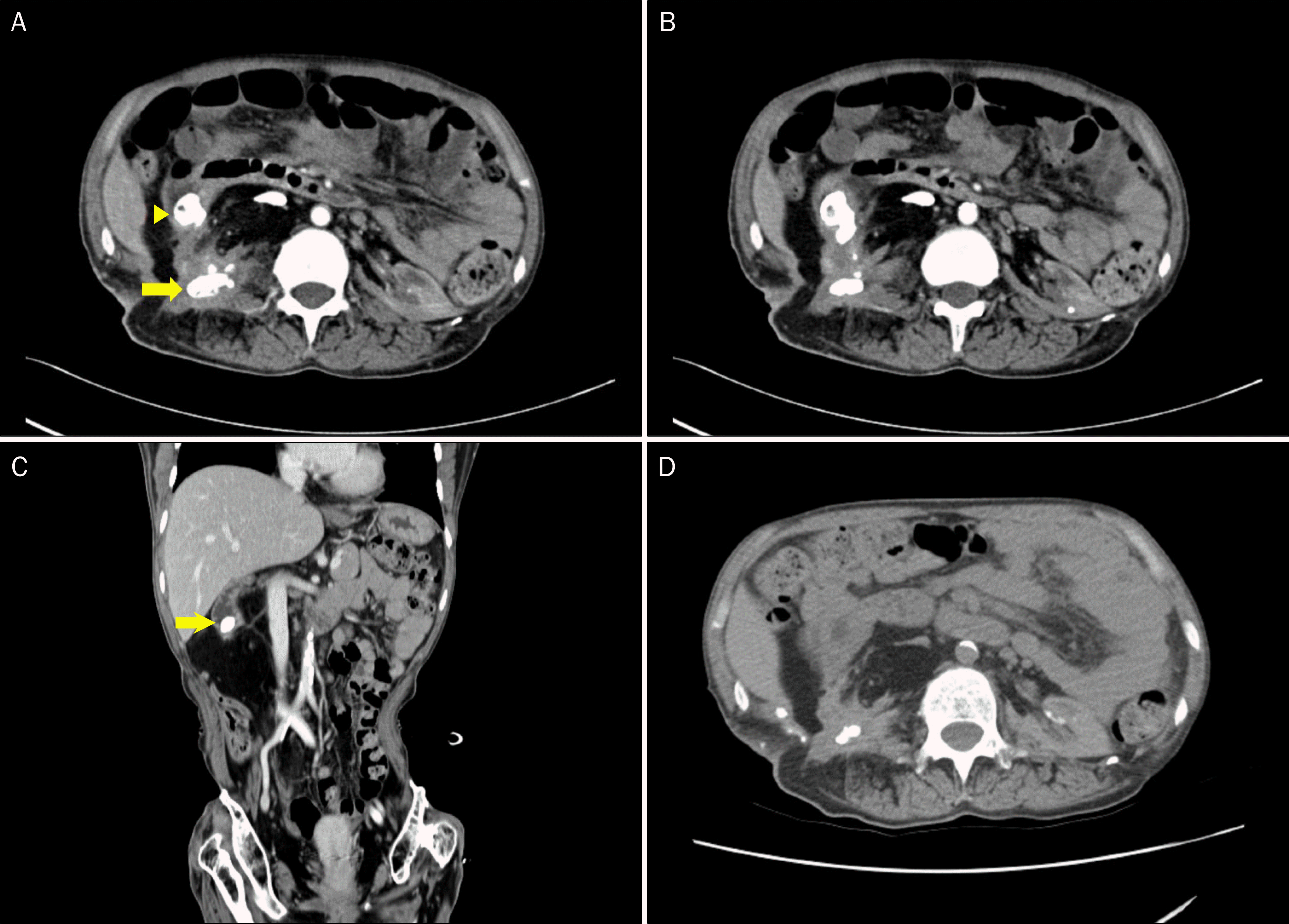
A fistula between the renal pelvis and duodenum (pyeloduodenal fistula) is very rare. It can occur spontaneously or after trauma to one of these organs. A spontaneous pyeloduodenal fistula is usually caused by chronic inflammation, including reactions to foreign bodies, nephrolithiasis, benign and malignant neoplasms, as well as pyogenic infections. The main treatment to date has been surgery. We encountered one case of pyeloduodenal fistula found during an evaluation for abdominal discomfort in a 39-year-old female. Pyeloduodenal fistula was diagnosed by upper gastrointestinal endoscopy and abdominal computed tomography, and it was caused by direct invasion of nephrolithiasis. Surgical operation was recommended, but the patient refused. The patient has been free of symptoms for four years. Herein, we report an unusual case of pyeloduodenal fistula without surgical management and relevant literature review.
Go to : 
References
1. Connor J, Schwartz M, Lehrhoff B. Nephrocolic fistula in association with a staghorn calculus discovered intraoperatively. Int Urol Nephrol. 1991; 23:113–116.

2. Hode E, Josse C, Mechaouri M, Garnier L, Verhaeghe P. Pyeloduodenal fistula. Apropos of a new case. J Chir (Paris). 1990; 127:281–285.
3. Abeshouse B. Renal and ureteral fistula of the visceral and cutaneous types; a report of four cases. Urol Cutaneous Rev. 1949; 53:641–674.
5. Lee KN, Hwang IH, Shin MJ, et al. Pyeloduodenal fistula successfully treated by endoscopic ligation without surgical nephrectomy: case report. J Korean Med Sci. 2014; 29:141–144.

6. Ginsberg DA, Stein JP, Grossfeld GD, Tarter T, Skinner DG. Traumatic pyeloduodenal fistula: a case report and review of the literature. Urology. 1996; 47:588–591.

7. Ross J, Tanna D. Pyeloduodenal fistula. J R Coll Surg Edinb. 1974; 19:51–53.
8. Morris DB, Siegelbaum MH, Pollack HM, Kendall AR, Gerber WL. Renoduodenal fistula in a patient with chronic nephrostomy drainage: a case report. J Urol. 1991; 146:835–837.

10. McElwee TB, Randall RD Jr, Meredith JH. Duodenoureteral fistula. South Med J. 1983; 76:1587–1588.

11. Batch AJ, Amery AH, Reddy ER. Pyeloduodenal fistula: a case report and review of the literature. Br J Surg. 1979; 66:31–34.

12. Morris SB, Knight MJ, Shearer RJ. Pyelo‐duodenal fistula: a new method of closure. Br J Urol. 1994; 73:464–465.

13. Suhler A, Schimmel F, Viville C. Intestinal urinary fistulas of renal and pelvis origin. Ann Urol (Paris). 1995; 29:8–10.
14. Tan SM, Teh CH, Tan PK. Duodeno-ureteric fistula secondary to chronic duodenal ulceration. Ann Acad Med Singapore. 1997; 26:850–851.
Go to : 
 | Fig. 1.Serial endoscopic findings. (A) At endoscopy 8 years ago, a shallow ulcerative lesion with nodular change and convergence of mucosal folds is observed at the second portion of the duodenum. (B) Endoscopy taken 2 years ago reveals an improvement of previous ulcerative lesion, but convergence of mucosal folds seems to still exist. (C) Endoscopy at visit reveals a brownish stone at the center of converging mucosal folds, protruding from the outside into the duodenal lumen. (D) Follow-up endoscopy 4 years later reveals a subsided stone and closed fistula. |
 | Fig. 2.Abdominal computed tomography. (A) In the right kidney, parenchymal calcification and staghorn calculi (arrow) are observed. The fragments of calculi invade into the duodenum (arrowhead). (B) There was a fistula between the second part of the duodenum and the right kidney. (C) On a coronal view, the fragments of calculi invading into the duodenum are clearly seen (arrow). (D) Follow-up computed tomography 4 years later reveals an improvement of calculi invasion of the duodenum. |


 PDF
PDF ePub
ePub Citation
 Print
Print


 XML Download