Abstract
Background/Aims
The clinical outcomes of angiography and transcatheter arterial embolization (TAE) for acute gastrointestinal bleeding (GIB) have not been completely assessed, especially according to bleeding sites. This study aimed to assess the efficacy of angiography and safety of TAE in acute GIB.
Methods
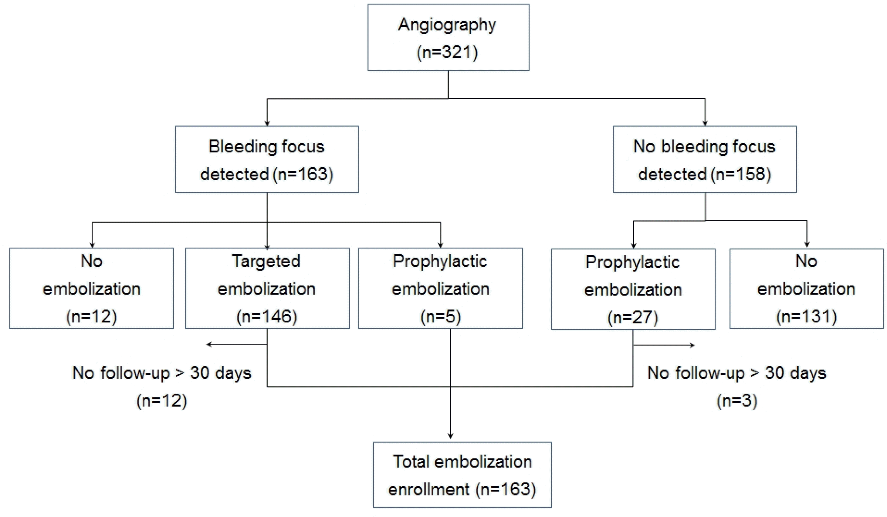
This was a retrospective study evaluating the records of 321 patients with acute GIB who underwent angiography with or without TAE. Targeted TAE was conducted in 134 patients, in whom angiography showed bleeding sources. Prophylactic TAE was performed in 29 patients when the bleeding source was not detected but a specific vessel was strongly suspected by other examinations. The rate of technical success, clinical success, and complications were analyzed.
Results
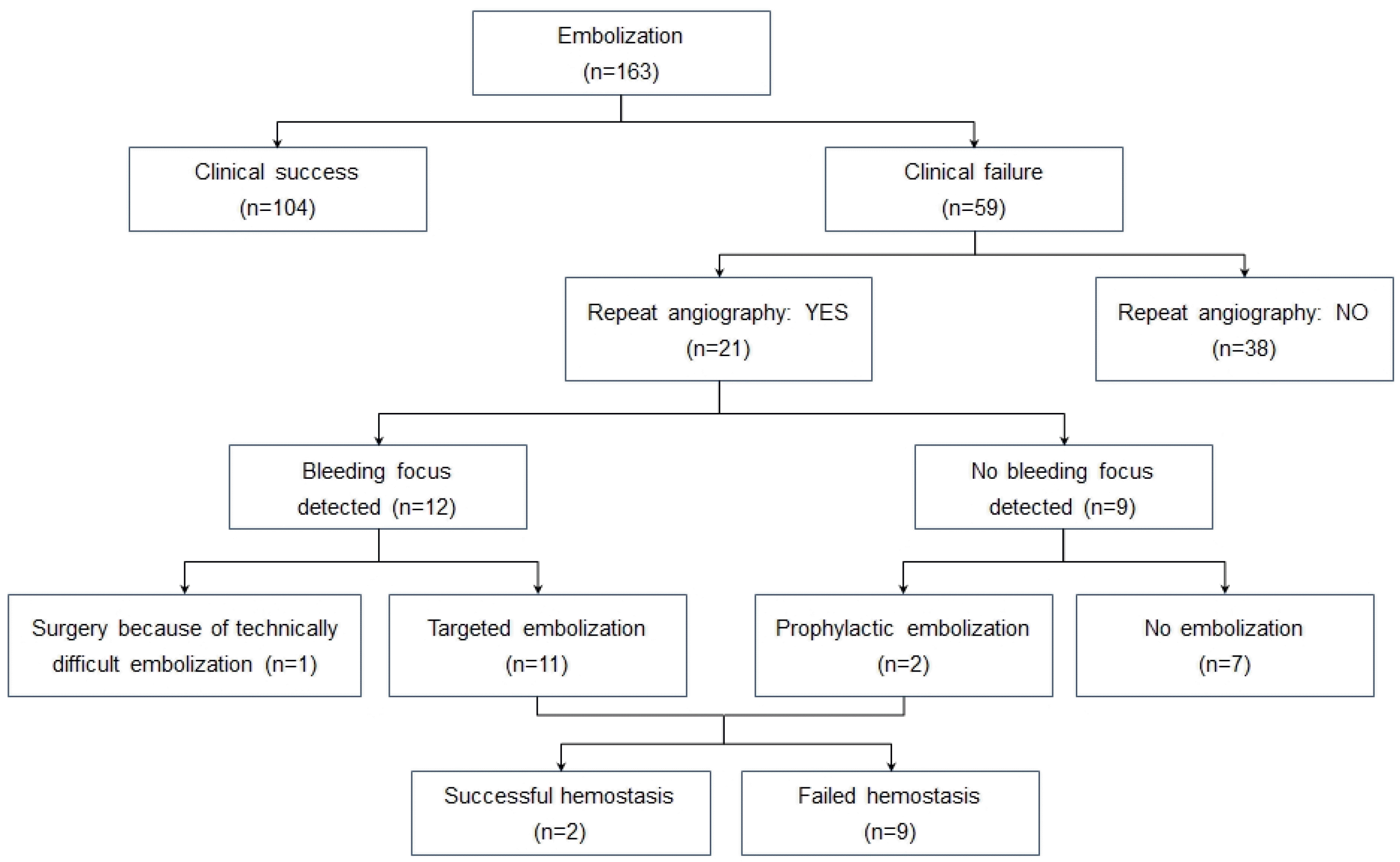
The detection rate of bleeding source via angiography was 50.8% (163/321), which was not different according to the bleeding sites. The detection rate was higher if the probable bleeding source had already been found by another investigation (59.7% vs. 35.8%, p<0.001). TAE sites were upper GIB in 67, mid GIB in 74, and lower GIB in 22. The technical success rate was 99.3% (133/134), and the clinical success rate was 63.0% (104/163). The prophylactic embolization group showed lower clinical success rate than the targeted embolization group (44.8% vs. 67.9%, p=0.06). The TAE-related complication rate was 12.9% (21/163). Ischemia and/or infarction was more common after TAE for mid and lower GIB than for upper GIB (15.6% vs. 3.0%, p=0.007).
Go to : 
References
1. Dye CE, Gaffney RR, Dykes TM, Moyer MT. Endoscopic and radiographic evaluation of the small bowel in 2012. Am J Med. 2012; 125:1228. e1–1228.e12.

2. Raju GS, Gerson L, Das A, Lewis B. American Gastroenterological Association. American Gastroenterological Association (AGA) institute medical position statement on obscure gastrointestinal bleeding. Gastroenterology. 2007; 133:1694–1696.

4. Barkun A, Bardou M, Marshall JK. Nonvariceal Upper GI Bleeding Consensus Conference Group. Consensus recommendations for managing patients with nonvariceal upper gastrointestinal bleeding. Ann Intern Med. 2003; 139:843–857.

5. Rollhauser C, Fleischer DE. Nonvariceal upper gastrointestinal bleeding. Endoscopy. 2004; 36:52–58.

6. Schenker MP, Duszak R Jr, Soulen MC, et al. Upper gastrointestinal hemorrhage and transcatheter embolotherapy: clinical and technical factors impacting success and survival. J Vasc Interv Radiol. 2001; 12:1263–1271.

7. Mirsadraee S, Tirukonda P, Nicholson A, Everett SM, McPherson SJ. Embolization for nonvariceal upper gastrointestinal tract haemorrhage: a systematic review. Clin Radiol. 2011; 66:500–509.

8. Lee IK, Kim YM, Kim J, et al. Superselective transarterial embolization for the management of acute gastrointestinal bleeding. J Korean Radiol Soc. 2006; 54:167–173.

9. Haglund U, Bergqvist D. Intestinal ischemia – the basics. Langenbecks Arch Surg. 1999; 384:233–238.

10. Dhatt HS, Behr SC, Miracle A, Wang ZJ, Yeh BM. Radiological evaluation of bowel ischemia. Radiol Clin North Am. 2015; 53:1241–1254.

12. Kwak HS, Han YM, Lee ST. The clinical outcomes of transcatheter microcoil embolization in patients with active lower gastrointestinal bleeding in the small bowel. Korean J Radiol. 2009; 10:391–397.

13. Maleux G, Roeflaer F, Heye S, et al. Longterm outcome of transcatheter embolotherapy for acute lower gastrointestinal hemorrhage. Am J Gastroenterol. 2009; 104:2042–2046.

14. Huang CC, Lee CW, Hsiao JK, et al. N-butyl cyanoacrylate embolization as the primary treatment of acute hemodynamically unstable lower gastrointestinal hemorrhage. J Vasc Interv Radiol. 2011; 22:1594–1599.

15. Kim BS, Li BT, Engel A, et al. Diagnosis of gastrointestinal bleeding: a practical guide for clinicians. World J Gastrointest Pathophysiol. 2014; 5:467–478.

16. Sildiroglu O, Muasher J, Arslan B, et al. Outcomes of patients with acute upper gastrointestinal nonvariceal hemorrhage referred to interventional radiology for potential embolotherapy. J Clin Gastroenterol. 2014; 48:687–692.

17. Pennoyer WP, Vignati PV, Cohen JL. Mesenteric angiography for lower gastrointestinal hemorrhage: are there predictors for a positive study? Dis Colon Rectum. 1997; 40:1014–1018.

18. Gunderman R, Leef J, Ong K, Reba R, Metz C. Scintigraphic screening prior to visceral arteriography in acute lower gastrointestinal bleeding. J Nucl Med. 1998; 39:1081–1083.
19. Steer ML, Silen W. Diagnostic procedures in gastrointestinal hemorrhage. N Engl J Med. 1983; 309:646–650.

20. Gerson LB, Fidler JL, Cave DR, Leighton JA. ACG clinical guideline: diagnosis and management of small bowel bleeding. Am J Gastroenterol. 2015; 110:1265–1287.

21. Okazaki M, Higashihara H, Koganemaru F, Ono H, Hoashi T, Kimura T. A coaxial catheter and steerable guidewire used to embolize branches of the splanchnic arteries. AJR Am J Roentgenol. 1990; 155:405–406.

22. Matsumoto AH, Suhocki PV, Barth KH. Superselective gelfoam embolotherapy using a highly visible small caliber catheter. Cardiovasc Intervent Radiol. 1988; 11:303–306.
23. Shin JH. Recent update of embolization of upper gastrointestinal tract bleeding. Korean J Radiol. 2012; 13(Suppl 1):S31–S39.

24. Holme JB, Nielsen DT, Funch-Jensen P, Mortensen FV. Transcatheter arterial embolization in patients with bleeding duodenal ulcer: an alternative to surgery. Acta Radiol. 2006; 47:244–247.

25. Larssen L, Moger T, Bjørnbeth BA, Lygren I, Kløw NE. Transcatheter arterial embolization in the management of bleeding duodenal ulcers: a 5.5-year retrospective study of treatment and outcome. Scand J Gastroenterol. 2008; 43:217–222.

26. Loffroy R, Guiu B, Cercueil JP, et al. Refractory bleeding from gastroduodenal ulcers: arterial embolization in high-operative-risk patients. J Clin Gastroenterol. 2008; 42:361–367.
27. Poultsides GA, Kim CJ, Orlando R 3rd, Peros G, Hallisey MJ, Vignati PV. Angiographic embolization for gastroduodenal hemorrhage: safety, efficacy, and predictors of outcome. Arch Surg. 2008; 143:457–461.
28. Loffroy R, Guiu B, D'Athis P, et al. Arterial embolotherapy for endoscopically unmanageable acute gastroduodenal hemorrhage: predictors of early rebleeding. Clin Gastroenterol Hepatol. 2009; 7:515–523.

29. Yap FY, Omene BO, Patel MN, et al. Transcatheter embolotherapy for gastrointestinal bleeding: a single center review of safety, efficacy, and clinical outcomes. Dig Dis Sci. 2013; 58:1976–1984.

30. Arrayeh E, Fidelman N, Gordon RL, et al. Transcatheter arterial embolization for upper gastrointestinal nonvariceal hemorrhage: is empiric embolization warranted? Cardiovasc Intervent Radiol. 2012; 35:1346–1354.

31. Ichiro I, Shushi H, Akihiko I, Yasuhiko I, Yasuyuki Y. Empiric transcatheter arterial embolization for massive bleeding from duodenal ulcers: efficacy and complications. J Vasc Interv Radiol. 2011; 22:911–916.

32. Loffroy R, Lin M, Thompson C, Harsha A, Rao P. A comparison of the results of arterial embolization for bleeding and nonbleeding gastroduodenal ulcers. Acta Radiol. 2011; 52:1076–1082.

33. Walker TG, Salazar GM, Waltman AC. Angiographic evaluation and management of acute gastrointestinal hemorrhage. World J Gastroenterol. 2012; 18:1191–1201.

34. Hongsakul K, Pakdeejit S, Tanutit P. Outcome and predictive factors of successful transarterial embolization for the treatment of acute gastrointestinal hemorrhage. Acta Radiol. 2014; 55:186–194.

35. Gillespie CJ, Sutherland AD, Mossop PJ, Woods RJ, Keck JO, Heriot AG. Mesenteric embolization for lower gastrointestinal bleeding. Dis Colon Rectum. 2010; 53:1258–1264.

36. Bua-Ngam C, Norasetsingh J, Treesit T, et al. Efficacy of emergency transarterial embolization in acute lower gastrointestinal bleeding: a single-center experience. Diagn Interv Imaging. 2017; 98:499–505.

Go to : 
Table 1.
Patient Demographic Data and Clinical Parameters (n=321)
| Variable | No. of patients |
|---|---|
| Male | 216 (67.3) |
| Age (yr) | |
| 18–60 | 154 (48.0) |
| 61–80 | 146 (45.5) |
| ≥81 | 21 (6.5) |
| Medications a | 82 (25.5) |
| Coagulopathy b | 87 (27.1) |
| Hemodynamic instability c | 165 (51.4) |
| Bleeding site | |
| UGI | 99 (30.8) |
| Peptic ulcer disease | 36 |
| Malignant | 22 |
| Acute gastroduodenal lesion | 6 |
| Dieulafoy's lesion | 4 |
| Others d | 17 |
| No source identified | 14 |
| MGI | 150 (46.7) |
| No source identified | 46 |
| Postoperative anastomotic bleedi | ng 19 |
| Malignant | 18 |
| Angioectasia | 17 |
| Inflammatory bowel disease | 15 |
| Others e | 35 |
| LGI | 48 (15.0) |
| Diverticulosis | 16 |
| Post operation bleeding | 4 |
| Angioectasia | 4 |
| Others f | 21 |
| No source identified | 3 |
| Unknown | 24 (7.5) |
UGI, upper gastrointestinal; MGI, mid gastrointestinal; LGI, lower gas-trointestinal; NSAIDs, nonsteroidal anti-inflammatory drugs; PT, prothrombin time; INR, international normalized ratio; CMV, cytomegalovirus.
c During or before the procedure, heart rate>120 beats per minute or systolic blood pressure <90 mmHg
d Iatrogenic, angiodysplagia, diverticular, fistula, Crohn's disease, vasculitis, esophageal hematoma, pseudoaneurysm, esophagitis
Table 2.
Indications and Diagnostic Yields of Angiography
| Angiography after detection of bleeding foc by other investigations (n=201) | Angiography with unknown bleeding focus (n=120) | p-value | |
|---|---|---|---|
| Diagnostic studies detecting bleeding focus | Upper and/or lower gastrointestinal | ||
| prior to angiography | endoscopy (n=90) | ||
| Abdominopelvic CT scan (n=37) | |||
| Bleeding scan (n=31) | |||
| CT enterography (n=25) | |||
| Enteroscopy (n=14) | |||
| Others a (n=4) | |||
| Diagnostic yield | |||
| Overall detection rate | 120 (59.7%) | 43 (35.8%) | <0.001 |
| UGI lesion detection rate | 39/74 (52.7%) | 14/25 (56.0%) | 0.775 |
| MGI lesion detection rate | 59/91 (64.8%) | 25/59 (42.4%) | 0.007 |
| LGI lesion detection rate | 22/33 (66.7%) | 4/15 (26.7%) | 0.014 |
Table 3.
Embolization Results according to the Bleeding Site
Table 4.
Summary of 21 Complications of Embolization
| No | Final diagnosis | Embolization vessel | Detection time a/diagnosis | Complication | Outcome |
|---|---|---|---|---|---|
| 1 | Gastric ulcer with Crohn's disease | Both GA | 6 hr/CT | Splenic infarction | Recovery & discharged |
| 2 | Dieulafoy's lesion below esophagojejunostomy ring | SMA br | 144 hr/EGD | Ischemic ulcer & bleeding | Recovery & discharged |
| 3 | Duodenal ulcer | GDA | 3 month/EGD | Duodenal stricture | Observation |
| 4 | Pancreatitis induced duodenal bleeding | GDA | 181 hr/EGD | Ischemic duodenitis | Recovery & discharged |
| 5 | Duodenal angiodysplasia | Both GA, left IPA | 14 hr/laboratory test | Acute kidney injury | Died of hyperkalemia after 4 days |
| 6 | SB bleeding | SMA br | 6 hr/clinically | Ischemic enteritis | Recovery & discharged |
| 7 | Radiotherapy related enteritis | SMA br | 55 hr/CT | Ischemic enteritis | Operation & discharged |
| 8 | SB traumatic bleeding | SMA br | 44 hr/colonoscopy | Ischemic enteritis | Recovery & discharged |
| 9 | SB angiodysplasia | SMA br | 21 hr/CT | Ischemic enteritis | Operation & discharged |
| 10 | SB bleeding | SMA br | 45 hr/CT | SB infarction with perforation | Operation & discharged |
| 11 | SB bleeding | SMA br | 27 hr/CT | Ischemic enteritis | Recovery & discharged |
| 12 | SB bleeding | SMA br | 66 hr/CT | Ischemic enteritis | Operation & successful hemostasis but died of leukemia |
| 13 | SB angiodysplasia | SMA br | 21 hr/CT | Ischemic enteritis | Recovery & discharged |
| 14 | SB bleeding | SMA br | 41 hr/CT | SB infarction with perforation | Operation & discharged |
| 15 | Splenic artery aneurysm | Splenic artery | 116 hr/CT | Splenic infarction | Recovery & discharged |
| 16 | Jejunostomy site bleeding | SMA br | 17 hr/CT | Ischemic enteritis | Operation & discharged |
| 17 | Jejunostomy site bleeding | SMA br | 179 hr/CT | SB infarction | Operation & discharged |
| 18 | Ascending colon diverticular bleeding | g SMA br | 551 hr/clinically | Ischemic colitis | Recovery & discharged |
| 19 | Post polypectomy bleeding | SMA br | 13 hr/CT | Ischemic colitis | Operation & discharged |
| 20 | Post polypectomy bleeding | SMA br | 16 hr/CT | Ischemic colitis | Recovery & discharged |
| 21 | Rectal AVM | IMA br | 61 hr/sigmoidoscopy | Ischemic colitis | Operation & discharged |


 PDF
PDF ePub
ePub Citation
 Print
Print


 XML Download