This article has been
cited by other articles in ScienceCentral.
Figures and Tables
Fig. 1
Endoscopic submucosal dissection process for treating an esophageal subepithelial tumor (SET). (A) A 2.5-cm esophageal SET located 23 cm from the central incisor. (B) An endoscopic ultrasound image showing a homogeneous hypoechoic tumor in the muscularis propria. (C) The covering mucosa of the SET stripped off by a coagulation snare. (D-F) The insulated-tip knife used to separate the tumor from the muscularis propria. (G) The tumor was completely separated from the esophageal wall. (H) Resected specimen, a 2.5×2 cm whitish tumor.
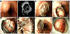
Fig. 2
Microscopic findings of a resected tumor. (A) Microscopically, the tumor was composed of irregularly oriented bundles of smooth muscle cells arranged in a whorl pattern (hemotoxylin and eosin, ×100). (B) Immunohistochemical stain (desmin, ×100) for desmin revealed positivity in the tumor cells.


 ePub
ePub Citation
 Print
Print


 XML Download