

 ePub
ePub Citation
 Print
Print


INTRODUCTION
Liver fibrosis and cirrhosis, the end-stage of hepatic fibrosis are the major causes of morbidity and mortality in patients with chronic liver disease.1 Activation of hepatic stellate cells (HSCs) in response to liver injury and the initiation of unbalanced synthesis of collagen are the main steps in the pathogenesis of fibrosis.2 Cirrhosis is characterized by architectural distortion of the liver, including diffuse parenchymal nodularity, fibrosis, and vascular changes in variable severity.3
Recently, it has been suggested that there is a significant relationship between gut microbiome and liver disease. Given that the liver is the first extraintestinal organ that encounters venous blood from the small and large intestines via the portal vein, it is most vulnerable to bacteria translocated form the gut lumen. Disruption of the intestinal epithelial barrier results in a leaky gut, which contributes to bacterial translocation. Consequently, translocated bacteria, such as lipopolysaccharides (LPSs), augment the activation of hepatic immune cells through pattern recognition receptors, including Toll-like receptors (TLRs). Moreover, hepatic non-immune cells, including endothelial cells, biliary epithelial cells, and hepatic stellate cells, also respond to bacterial products through TLRs.4567
Rifaximin is a non-absorbable antibiotic used for intestinal decontamination. It can be an effective treatment for subclinical and overt hepatic encephalopathy. It was reported to reduce endotoxemia and severity of liver disease in humans.8 In a recent animal study, rifaximin was reported to inhibit TLR4 dependent fibronectin mediated crosstalk, between stellate cells and endothelial cells in liver fibrosis in a 21-day bile duct-ligated mice model.9 However, inflammation and fibrosis were not simultaneously investigated. Those were not explored in the early stages of hepatic fibrosis on rifaximin treatment.
Therefore, in this study, we investigated the effects of rifaximin on hepatic inflammation and fibrosis in a bile duct-ligated rat model of acute and chronic liver injury.
SUBJECTS AND METHODS
1. Materials and animal experiments
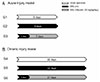
Rifaximin was provided by Hanall Biopharma (Seoul, Korea). Male adult Sprague-Dawley rats, weighing 200-250 g, were purchased from Orient Bio Inc. (Seongnam, Korea). The rats were maintained in a temperature-controlled room (22±2℃), with 12 hours light/dark cycle and 40-60% humidity, with ad libitum access to food and water. Bile duct ligation (BDL) was performed for 8 and 22 days in the acute and chronic injury models, respectively. Some rats were divided into three groups (for 8 days): sham-operated rats (sham G1 [n=6]), BDL rats (BDL G2 [n=4]), and BDL rats treated with rifaximin (BDL-rifaximin G3 [n=9]). Similarly, some rats were allocated to three groups in the chronic injury model (for 22 days): sham-operated rats (sham G4 [n=7]), BDL rats (BDL G5 [n=6]), and BDL rats treated with rifaximin (BDL-rifaximin G6 [n=5]). After BDL, rifaximin (50 mg/kg/day) was administered daily via gavage for 7 days in the case of acute injury model and 21 days in the case of chronic injury model (Fig. 1).
2. Bile duct ligation
For all surgical procedures, the rats were anesthetized by inhalation of 5 vol% isoflurane in 100% oxygen at a flow rate of 4 L/min for the induction and 2-3 vol% at 2 L/min for the maintenance. In the sham operated group, a midline incision in the abdomen was made using the sterile techniques. In the BDL and BDL-rifaximin groups, after a midline laparotomy of 1-2 cm, the common bile duct was identified, doubly ligated using 5-0 silk sutures, and transected at the level of 0.7-0.8 cm distal to the last bifurcation. The first ligature was made below the junction of the hepatic ducts, and the second ligature was made above the entrance to the pancreatic ducts. Finally, the common bile duct was resected between the double ligatures. Each animal received a subcutaneous injection of 10 mg/kg ketoprofen for 1 day and 5 mg/kg gentamicin for 3 days, after the surgery. Rats in the sham G1, BDL G2, and BDL-rifaximin G3 groups were sacrificed on day 8, whereas those in the sham G4, BDL G5, and BDL-rifaximin G6 groups were sacrificed on day 22, under isoflurane anesthesia. Serum samples were collected and stored at -80℃. Freshly dissected liver samples were fixed, processed, and paraffin-embedded for histologic examination. Liver tissues were snap-frozen in liquid nitrogen and stored at -80℃ for further analysis. All animal experiments were approved by the Gachon University Institutional Animal Care and Use Committee (LCDI-2015-0009).
3. Serum biochemistry
Serum total bilirubin, alanine aminotransferase (ALT), and aspartate aminotransferase (AST) levels were analyzed with an automatic analyzer (cobas c111 system; Roche Diagnostics Ltd., Rotkreuz, Switzerland).
4. Determination of serum tumor necrosis factor-alpha (TNF-α) level
Serum TNF-α levels were measured using the commercial enzyme linked immunosorbent assay kit (R&D systems, Minneapolis, MN, USA) according to the manufacturer's instructions. The absorbance was recorded using an EMax® microplate reader (Molecular Devices, Sunnyvale, CA, USA), and concentrations were calculated based on the standard curve.
5. Hepatic hydroxyproline content
Hepatic hydroxyproline levels were quantified in accordance with the manufacturer's protocol using the colorimetric hydroxyproline assay kit (Biovision, Mountview, CA, USA). Liver tissue (10 mg) was incubated with 100 µL of 12 N HCl for 3 hours at 120℃. The hydrolyzed sample (10 µL) was transferred to a 96-well plate and allowed to dry. Chloramine-T (100 µL) was added to each well and the plate was incubated at 60℃ for 90 minutes. The absorbance at 560 nm was recorded using an EMax® microplate reader (Molecular Devices, Sunnyvale, CA, USA), and concentrations were calculated based on the standard curve.
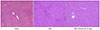
6. Histologic evaluation
Three-micrometer-thick sections obtained from the paraffin-embedded livers were stained with a hematoxylin and eosin (H&E) solution to evaluate structural changes. Paraffin-embedded tissue sections were also stained with picrosirius red (Sigma-Aldrich, St. Louis, MO, USA) and counterstained with fast green (Sigma-Aldrich, St. Louis, MO, USA) using previously published methods for the quantification of liver fibrosis content.10 Histological images of each sample were analyzed from six random 100× fields using Image J software (National Institutes of Health, Bethesda, MD, USA), and the percentage of red-stained area, which is a marker for collagen content, was measured. The data were pooled to determine the mean.
RESULTS
1. Effect of rifaximin on serum biochemistry in acute or chronic injury model
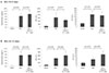
The total bilirubin levels were higher in BDL (BDL G2 and G5) and BDL-rifaximin groups (BDL-rifaximin G3 and G6) than in the sham groups (sham G1 and G4). However, the total bilirubin levels were not different between the BDL and BDL-rifaximin groups. Similarly, the serum AST and ALT levels were also higher in the BDL (BDL G2 and G5) and BDL-rifaximin groups (BDL-rifaximin G3 and G6) than in the sham groups (sham G1 and G4). However, the serum AST levels were not different between the BDL and BDL-rifaximin groups. In the case of serum ALT levels, there was no difference between BDL G2 and BDL-rifaximin G3. Moreover, the serum ALT level was rather higher in BDL-rifaximin G6 than in BDL G5 (Fig. 2).
2. Effect of rifaximin on serum TNF-α level in acute or chronic injury model
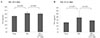
The serum TNF-α levels (pg/well) in sham G1, BDL G2, and BDL-rifaximin G3 were 22.5±1.1, 26.5±2.1, and 25.8±1.5 respectively. The level was significantly elevated in BDL G2 compared with that in sham G1. Although there was a decrease in serum TNF-α level in BDL-rifaximin G3 compared with that in BDL G2, the difference was not statistically significant (Fig. 3A). Serum TNF-α levels (pg/well) in sham G4, BDL G5, and BDL-rifaximin G6 were 16.1±0.4, 24.2±10.8, and 19.2±1.3 respectively. The level was not significantly elevated in BDL G5 compared with that in sham G4 (p=0.074). Although there was a decrease in the serum TNF-α level in BDL-rifaximin G6 compared with that in BDL G5, the difference was not statistically significant (Fig. 3B).
3. Effect of rifaximin on hydroxyproline level in acute or chronic injury model
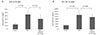
The hydroxyproline levels (µg/g liver tissue) in sham G1, BDL G2, and BDL-rifaximin G3 were 236.4±103.1, 444.8± 114.4, and 312.5±131.6, respectively. The level was significantly higher in BDL G2 than in sham G1 (p=0.010). Although the level was lower in BDL-rifaximin G3 than in BDL G2, rifaximin treatment did not lead to a statistically significant reduction in the hydroxyproline level (Fig. 4A). The hydroxyproline levels in sham G4, BDL G5, and BDL-rifaximin G6 were 311.5±72.9, 1,110.3±357.9, and 944.3±209.3, respectively. The level was significantly higher in BDL G5 than in sham G4 (p<0.001). Although the level was lower in BDL-rifaximin G6 than in BDL G5, rifaximin treatment did not lead to statistically significant reduction in the level of hydroxyproline (Fig. 4B).
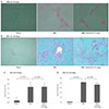
4. Effect of rifaximin on fibrosis content in acute or chronic injury model
H&E staining of the liver from chronic injury model showed an increased inflammatory cell infiltration with bridging necrosis and fibrotic band interposition. H&E staining did not reveal any histological improvement after the rifaximin treatment (Fig. 5). After sirius red staining, remarkable collagen accumulation was observed in the BDL groups, based on the hyperplasia of the lattice fibers and collagenous fibers in the portal area. In both acute and chronic injury models, collagen accumulation measured by sirius red staining in the BDL-rifaximin groups was not different from that in the BDL groups (Fig. 6A, B). In the acute injury model, collagen contents (%) in sham G1, BDL G2, and BDL-rifaximin G3 were 0.22±0.04, 1.64±0.53, and 1.66±0.44, respectively. While the fibrosis content was significantly higher in BDL G2 than in sham G1 (p=0.001), there was no difference between BDL G2 and BDL-rifaximin G3 (Fig. 6C). In the chronic injury model, the collagen contents (%) in sham G4, BDL G5, and BDL-rifaximin G6 were 0.19±0.03, 5.04±0.18, and 4.42±0.68, respectively. While the fibrosis content was significantly higher in BDL G5 than in sham G4 (p<0.001), it was marginally decreased by the rifaximin treatment in BDL-rifaximin G6 compared with that in BDL G5 (p=0.059) (Fig. 6D).
DISCUSSION
Ligation of the common bile duct is one of the most common methods for inducing liver fibrosis. Biliary obstruction by BDL can lead to hepatocellular injury, followed by bile duct proliferation, fibrosis, and ultimately cirrhosis. Depending on the time of obstruction, it can result in either acute or chronic hepatic damage.11 A previous study using a bile duct-ligated rat model showed that the serum AST and ALT levels peaked in the 1st week after surgery, and decreased in the 2nd week.11 The present study also showed that the serum AST and ALT levels in the acute injury model (8-day bile duct-ligated rat model) were higher than those in the chronic injury model (22-day bile duct-ligated rat model). Zhu et al.9 reported that rifaximin treatment did not lead to a statistically significant improvement in liver function after BDL. This was substantiated in the present study, as rifaximin treatment did not improve liver function in both acute and chronic injury models. These results indicate that rifaximin treatment cannot ameliorate the hepatocellular injury caused by BDL.
Kupffer cells (KCs) play a key role in modulating the inflammatory response observed in most animal models of liver injury.12 LPS-induced activation of KCs in animal models leads to the secretion of a number of pro-inflammatory cytokines, such as TNF-α, interleukin 1 (IL-1), and IL-6.13 Contrastingly, in response to LPS stimulation, freshly isolated human KCs secrete anti-inflammatory cytokine IL-10, which contributes to the downregulation of pro-inflammatory cytokines, and possibly, improved LPS tolerance of KCs.14 Moreover, a previous study reported that the serum TNF-α level on the 14th day in the BDL group was not highly elevated compared with that in the controls, although it was highly elevated on the 7th day after BDL.15 In the present study, the serum TNF-α level was elevated in the acute injury model than in the sham group after BDL. However, it was not significantly elevated in the chronic injury model when compared with that in the sham group after BDL. If the LPS levels were decreased by rifaximin treatment, the serum TNF-α level is expected to decrease. However, such an effect was not observed after rifaximin treatment in the present study. There was a limitation in confirming the effects of rifaximin on anti-inflammation. Thus, further studies are necessary to detect other inflammatory cytokine pathways that is influenced by rifaximin treatment. A previous report has shown that HSCs, and not KCs, are the primary targets driving fibrogenesis in response to TLR4 ligands. The same report also implicated TLR4 expressed on HSCs as the key driver of liver fibrosis owing to the stimulatory effects of TLR4 activation on the transforming growth factor-beta pathway.16 Seki et al.16 demonstrated that the intestinal microflora, which is the main source of portal LPS and ligands for TLR4 activation, is an important prerequisite for the development of liver fibrosis after BDL. After BDL, TLR4-wild-type mice showed overt hepatic fibrosis, whereas TLR4-mutant mice had a significant reduction in fibrosis as demonstrated by sirius red staining, hydroxyproline levels, and α-smooth muscle actin expression.
Zhu et al.9 demonstrated that rifaximin treatment for 3 weeks attenuated BDL induced fibronectin formation and fibrosis, which were confirmed by immunofluorescent staining of fibronectin and histopathological changes observed after sirius red staining, respectively. However, the acute injury model in this present study showed that the hydroxyproline levels and quantitation of fibrosis using sirius red staining were not decreased by rifaximin treatment compared with the BDL group. This result showed that the duration of the treatment was too short for rifaximin to fully exert its anti-fibrotic activity.
While the hydroxyproline levels were not decreased by rifaximin treatment in our chronic injury model, the fibrosis contents, as measured by sirius red staining, were marginally decreased. These contradictory results might be due to the use of different techniques for the quantification of fibrosis content. While the hydroxyproline assay involved the use of small pieces of liver tissue, instead of the whole liver, the fibrosis content analysis via sirius red staining can be performed on a much larger area of the liver. Compared with a previous mouse study by Zhu et al.,9 the anti-fibrotic effect of rifaximin was not significant in this rat study. Difference of subject animal may influence different results. However, the rifaximin dosage used in this study was commonly used in other rat studies.1718 In addition, it seems that rifaximin administration for 3 weeks was not enough to exhibit adequate anti-fibrotic action. Although rifaximin has not been extensively investigated with appropriate duration for its full anti-fibrotic effect, a previous study has shown a decrease in hepatic venous pressure gradient in patients with alcohol-related decompensated cirrhosis after 4 weeks of rifaximin administration.19 Therefore, studies for long-term treatment of rifaximin―greater than 4 weeks in duration―are warranted. After all, our results showed that liver fibrosis in bile duct-ligated rat models was not only predominantly LPS-induced. Therefore, rifaximin treatment, which mainly eradicates intestinal bacteria, may not completely prevent the development of liver fibrosis on its own.
Our study has several limitations to consider. First, the levels of intestinal microflora were not measured before and after the rifaximin treatment. Second, LPS levels were not measured, because the rifaximin treatment did not significantly affect the TNF-α level and fibrosis content. Third, other liver fibrosis models using hepatic toxins, such as carbon tetrachloride, thioacetamide, and diethylnitrosamine, were not performed in our study. To elucidate more precise effects and mechanisms of rifaximin, further studies using various models of liver fibrosis will be necessary.
In conclusion, the short- and long-term effects of rifaximin treatment were unsatisfactory with respect to the inhibition of inflammation and fibrosis in bile duct-ligated rat models. Although rifaximin only marginally inhibited liver fibrosis, the main mechanism behind liver fibrosis formation in bile duct-ligated rat models might not be LPS-dependent. If a specific LPS-induced inflammation and fibrosis model (rather than a bile duct ligated model) is used, rifaxmin treatment may significantly inhibit inflammation and fibrosis. Additional studies on the effects of rifaximin treatment on other LPS-associated complications of liver cirrhosis are needed.
 XML Download