Abstract
Background/Aims
In recent years, the incidence of acute pancreatitis (AP) has been increasing. A better understanding of the etiology is directly linked to more favorable outcomes. Unfortunately, there have been reports suggesting the variation of etiologies of AP across countries. The objective of this study was to determine the etiology of AP in a general hospital of Seoul-Gyeonggi province in Korea during the past decade.
Methods
We retrospectively reviewed the medical records of consecutive patients with AP who were admitted to St. Paul's Hospital (Seoul, Korea) with an affiliation to the Catholic University of Korea between January 2003 and January 2013.
Results
A total of 1,110 patients were enrolled, totaling 1,833 attacks, and the most frequent cause of AP was alcohol consumption. The recurrence rate of AP was 24.5% (272/1,110), and habitual recurrence rate (more than three times) was 12.6% (140/1,110). The rate of severe AP was 4.9% (90/1,833 attacks). The mortality rate of AP was 2.6% (29/1,110 patients). The frequency of an idiopathic cause of AP was 13.3%. The recurrence rate and mortality rate of idiopathic AP were 16.2% and 5.4%, respectively. In 41.7% (10/24) of cases of idiopathic AP, microlithiasis was suspected.
References
1. Cavallini G, Frulloni L, Bassi C, et al. Prospective multicentre survey on acute pancreatitis in Italy (ProInf-AISP): results on 1005 patients. Dig Liver Dis. 2004; 36:205–211.

2. Kim CD. Current status of acute pancreatitis in Korea. Korean J Gastroenterol. 2003; 42:1–11.
3. Hamada S, Masamune A, Kikuta K, et al. Nationwide epidemiological survey of acute pancreatitis in Japan. Pancreas. 2014; 43:1244–1248.

4. Lim YS, Ryu JK, Lee HC, Kim YT, Yoon YB, Kim CY. Comparison of etiological and prognostic factors in acute nexrcotizing pancreatitis. Korean J Gastroenterol. 1997; 29:667–676.
5. Sandzén B, Rosenmuller M, Haapamäki MM, Nilsson E, Stenlund HC, Oman M. First attack of acute pancreatitis in Sweden 1988 – 2003: incidence, aetiological classification, procedures and mortality – a register study. BMC Gastroenterol. 2009; 9:18.

6. Omdal T, Dale J, Lie SA, Iversen KB, Flaatten H, Ovrebo K. Time trends in incidence, etiology, and case fatality rate of the first attack of acute pancreatitis. Scand J Gastroenterol. 2011; 46:1389–1398.

7. van Brummelen SE, Venneman NG, van Erpecum KJ, VanBerge- Henegouwen GP. Acute idiopathic pancreatitis: does it really exist or is it a myth? Scand J Gastroenterol Suppl. 2003; (239):117–122.
8. Nordback I, Pelli H, Lappalainen-Lehto R, Järvinen S, Räty S, Sand J. The recurrence of acute alcohol-associated pancreatitis can be reduced: a randomized controlled trial. Gastroenterology. 2009; 136:848–855.

9. Gentilello LM, Ebel BE, Wickizer TM, Salkever DS, Rivara FP. Alcohol interventions for trauma patients treated in emergency departments and hospitals: a cost benefit analysis. Ann Surg. 2005; 241:541–550.
10. Yadav D, O'Connell M, Papachristou GI. Natural history following the first attack of acute pancreatitis. Am J Gastroenterol. 2012; 107:1096–1103.

11. Lankisch PG, Breuer N, Bruns A, Weber-Dany B, Lowenfels AB, Maisonneuve P. Natural history of acute pancreatitis: a long-term population-based study. Am J Gastroenterol. 2009; 104:2797–2805. quiz 2806.

12. Gullo L, Migliori M, Oláh A, et al. Acute pancreatitis in five European countries: etiology and mortality. Pancreas. 2002; 24:223–227.

14. Kim KP, Kim MH, Kim JC, Lee SS, Seo DW, Lee SK. Diagnostic criteria for autoimmune chronic pancreatitis revisited. World J Gastroenterol. 2006; 12:2487–2496.

15. Naranjo CA, Shear NH, Lanctôt KL. Advances in the diagnosis of adverse drug reactions. J Clin Pharmacol. 1992; 32:897–904.

16. Güngör B, Cağ layan K, Polat C, Seren D, Erzurumlu K, Malazgirt Z. The predictivity of serum biochemical markers in acute biliary pancreatitis. ISRN Gastroenterol. 2011; 2011:279607.
17. Grau F, Almela P, Aparisi L, et al. Usefulness of alanine and aspartate aminotransferases in the diagnosis of microlithiasis in idiopathic acute pancreatitis. Int J Pancreatol. 1999; 25:107–111.

18. Stimac D, Lenac T, Marusić Z. A scoring system for early differentiation of the etiology of acute pancreatitis. Scand J Gastroenterol. 1998; 33:209–211.
19. Tenner S, Dubner H, Steinberg W. Predicting gallstone pancreatitis with laboratory parameters: a metaanalysis. Am J Gastroenterol. 1994; 89:1863–1866.
20. Davidson BR, Neoptolemos JP, Leese T, Carr-Locke DL. Biochemical prediction of gallstones in acute pancreatitis: a prospective study of three systems. Br J Surg. 1988; 75:213–215.

21. World Health Organization. Global status report on alcohol and health. 1st ed.Geneva: World Health Organization;2011.
22. Samokhvalov AV, Rehm J, Roerecke M. Alcohol consumption as a risk factor for acute and chronic pancreatitis: a systematic review and a series of meta-analyses. EBioMedicine. 2015; 2:1996–2002.

23. Kume K, Masamune A, Ariga H, Shimosegawa T. Alcohol consumption and the risk for developing pancreatitis: a case-control study in Japan. Pancreas. 2015; 44:53–58.
24. Roberts SE, Akbari A, Thorne K, Atkinson M, Evans PA. The incidence of acute pancreatitis: impact of social deprivation, alcohol consumption, seasonal and demographic factors. Aliment Pharmacol Ther. 2013; 38:539–548.

25. O'Farrell A, Allwright S, Toomey D, Bedford D, Conlon K. Hospital admission for acute pancreatitis in the Irish population, 1997 2004: could the increase be due to an increase in alcohol-related pancreatitis? J Public Health (Oxf). 2007; 29:398–404.
26. Stevens CL, Abbas SM, Watters DA. How does cholecystectomy influence recurrence of idiopathic acute pancreatitis? J Gastrointest Surg. 2016; 20:1997–2001.

27. Del Chiaro M, Verbeke CS, Kartalis N, et al. Short-term results of a magnetic resonance imaging-based Swedish screening program for individuals at risk for pancreatic cancer. JAMA Surg. 2015; 150:512–518.

Fig. 1.
Etiology of acute pancreatitis in Korea. Alcohol consumption and gallstones are the main etiologic factors of AP. Other etiology of AP includes autoimmune (n=14), drug-induced (n=7), cancer-related (n=15), IPMN (n=8), pancreas divisum (n=5), and hypertriglyceridemia (n=4). AP, acute pancreatitis; IPMN, intraductal papillary mucinous neoplasm.
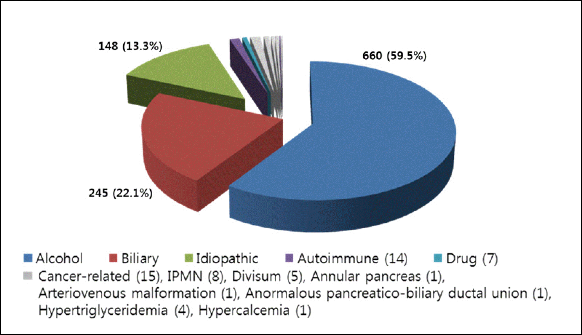
Fig. 2.
Autoimmune pancreatitis. (A) Pancreatic parenchymal swelling (arrow) and peripancreatic lower density rim like change (arrowhead). (B) Focal low-attenuating mass (arrow) around the celiac axis and adjacent para-aortic space in a patient with retroperitoneal fibrosis.
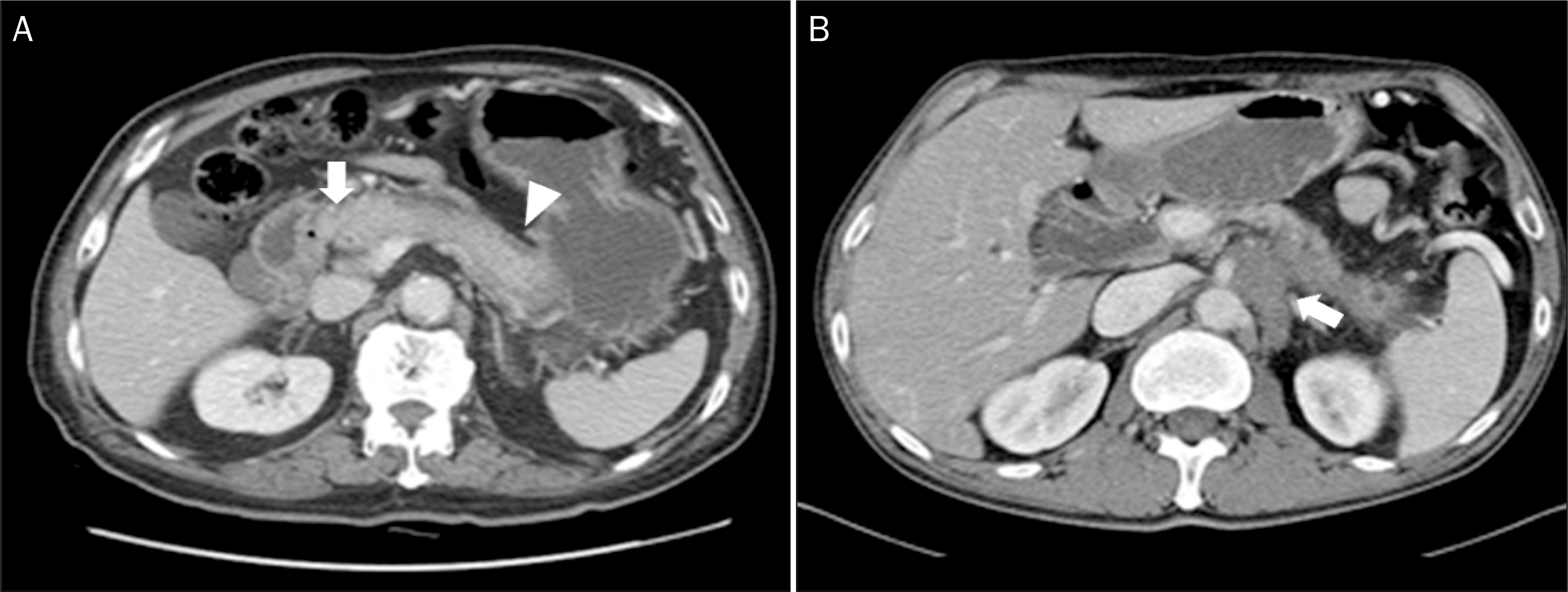
Fig. 3.
Unusual cause of acute pancreatitis-APBDU. (A) In MRCP image, fusiform dilatation of extrahepatic duct (arrow) suggestive of choledochal cyst is observed (Todani classification IA). (B). Fluoroscopic image taken during ERCP showing long common channel (arrow), a definitive finding of APBDU. APBDU, anomalous pancreaticobiliary ductal union; MRCP, magnetic resonance cholangiopancreatography; ERCP, endoscopic retrograde cholangiopancreatography.
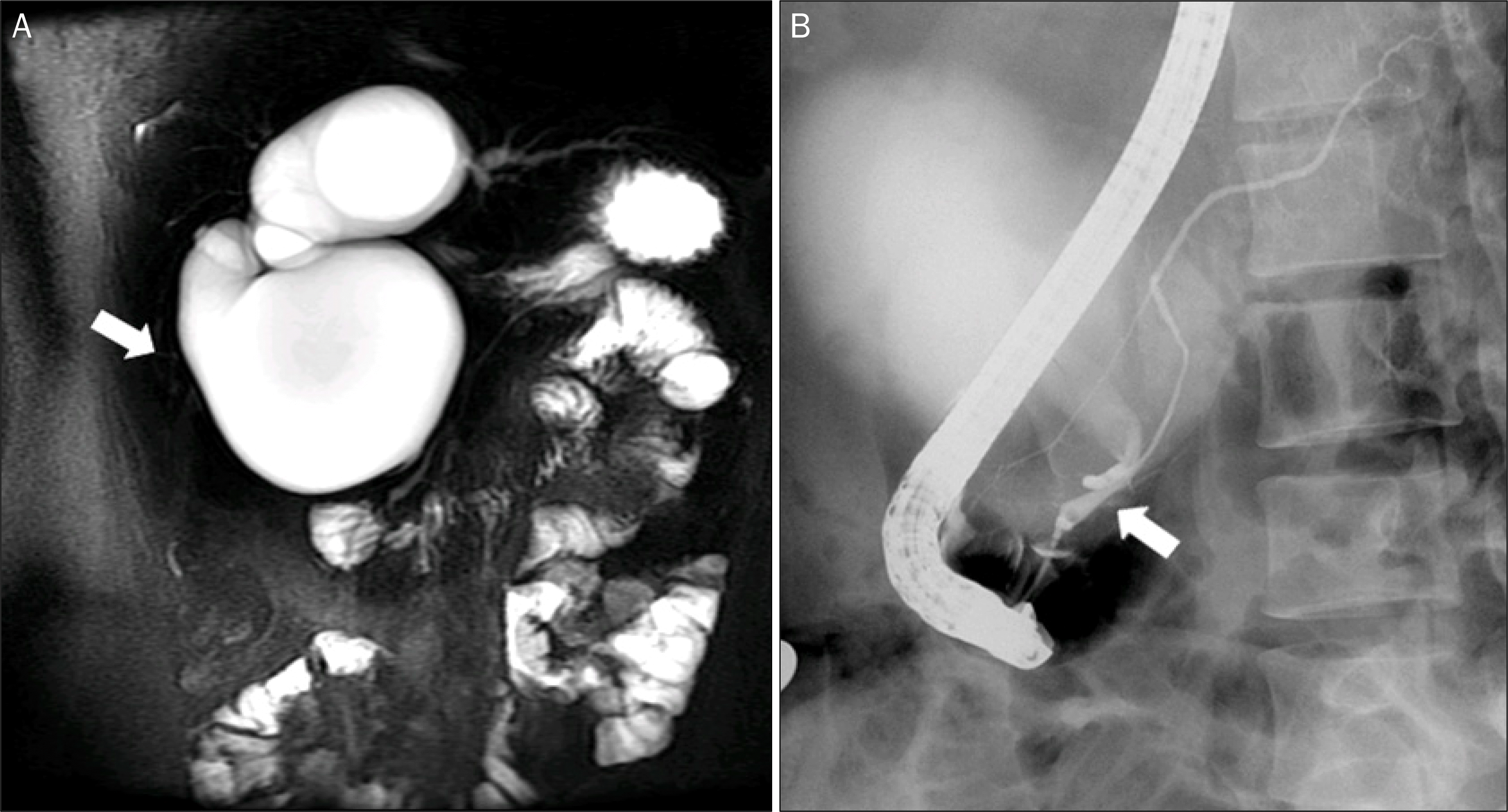
Fig. 4.
Unusual cause of acute pancreatitis-pancreatic AVM. (A) Acute intrapancreatic parenchymal bleeding and hematoma formation (arrow) is observed on CT coronal image. (B) In celiac artery angiography, dense stain in the pancreatic head region and early filling of the veins (double arrows) suggests pancreatic AVM. AVM, arteriovenous malformation; CT, computed tomography.
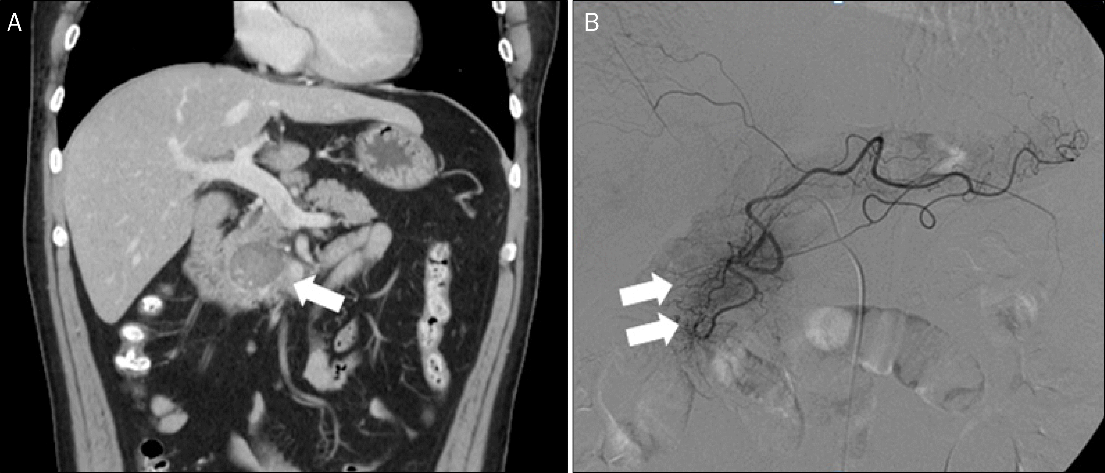
Fig. 5.
Unusual cause of acute pancreatitis-pancreatic cancer. (A) Fluoroscopic image taken during ERCP shows focal segmental pancreatic duct narrowing (double arrows) in the proximal body area with post-stenotic dilatation. (B) About 3.0 cm sized ill-defined mass (arrow) of the proximal body of pancreas and pancreatic duct dilatation (arrowhead) of the tail is observed in CT axial view. ERCP, endoscopic retrograde cholangiopancreatography; CT, computed tomography.
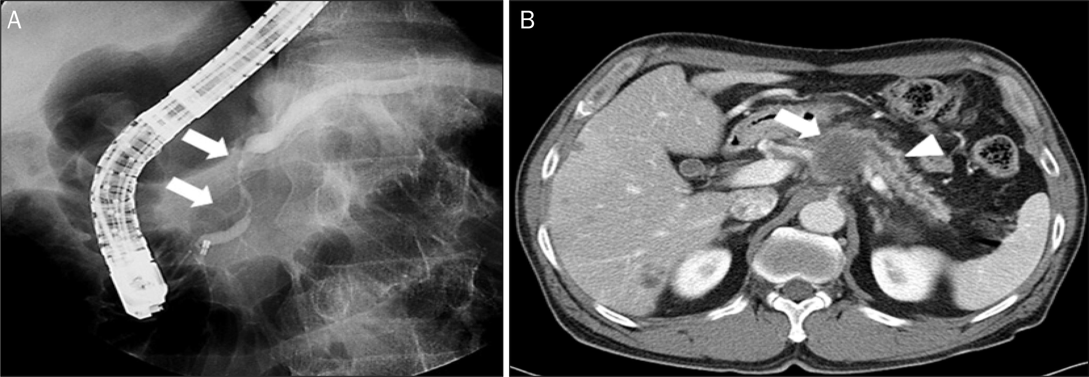
Fig. 6.
Unusual cause of acute pancreatitis-non-Hodgkin's lymphoma and lung cancer. (A) Peripancreatic mass invading the adjacent pancreatic body parenchyme (arrow) resulting in pancreatitis in the pancreatic tail (arrowhead) in diffuse large B cell lymphoma patient. (B) Heterogeneous contrast enhancing mass in the pancreatic tail (arrow) with peri-pancreatic infiltrations in lung cancer patient, suggesting pancreatic metastasis.
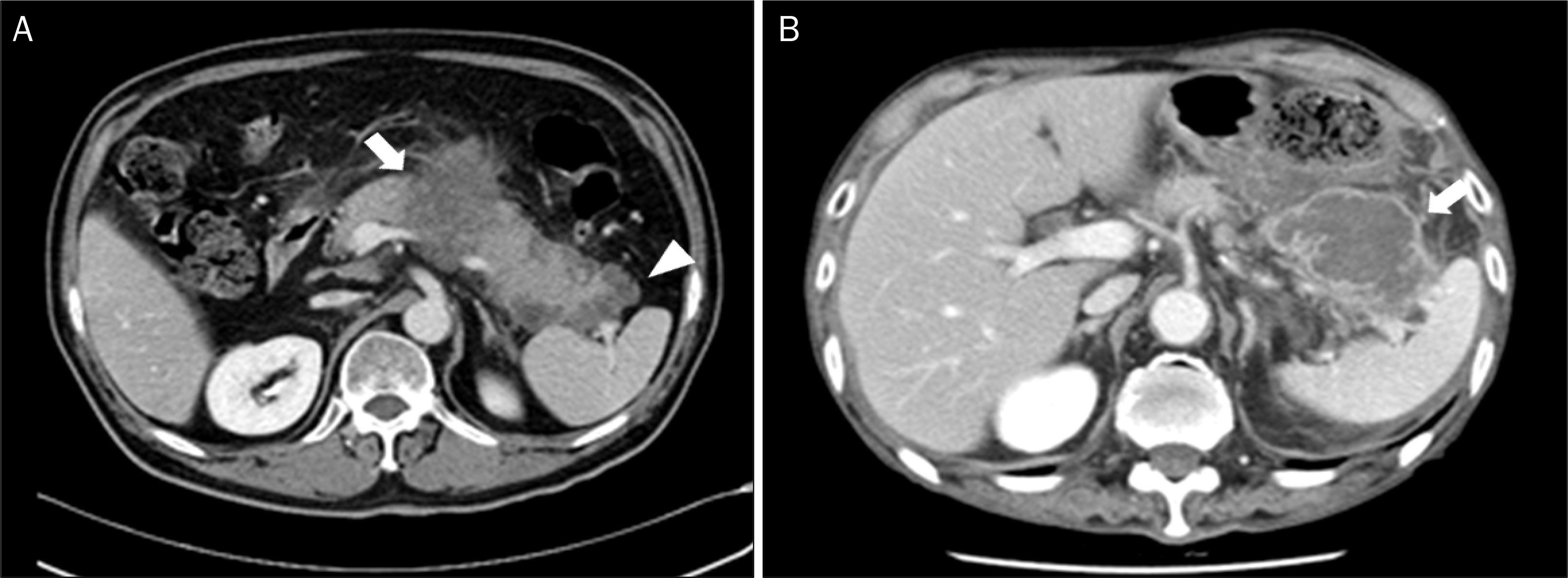
Table 1.
Etiologic Analysis of Acute Pancreatitis in Korea


 PDF
PDF ePub
ePub Citation
 Print
Print


 XML Download