Journal List > Korean J Gastroenterol > v.70(4) > 1007695

Kim, Lee, Kwon, Lee, Kim, Sung, Park, and Shim: Serological Changes after Equivocal Helicobacter pylori-serology Test Findings Dependent on Gastric Secreting Ability

Abstract

Background/Aims

The serum anti-Helicobacter pylori (H. pylori) immunoglobulin G (IgG) and serum pepsinogen (PG) assays are widely used to screen for gastric cancer. An equivocal serology test finding indicates IgG titer between the positive and negative test findings. This study aims to evaluate the long-term follow-up result after an equivocal test finding on the serum anti-H. pylori IgG assay.

Methods

Koreans aged 18 years or older with an equivocal serum anti-H. pylori IgG assay finding were included. Subjects were excluded if they did not undergo H. pylori serology test, serum PG assay, and upper gastrointestinal (UGI) endoscopy on the same day at our center. The annual test findings were followed-up using the same methods.

Results

Of the 7,178 subjects who underwent the serum assays and UGI endoscopy on the same day, 274 (3.8%) subjects showed an equivocal H. pylori serology test finding. Of the 98 subjects who were followed-up, 58 (59.2%) showed seropositive finding at the mean follow-up period of 30.6±12.4 months. Subjects with seroconversion showed a higher initial serum PG I (p=0.023) and PG II (p=0.036) levels than those without seroconversion.

Conclusions

An equivocal H. pylori serology test finding was not rare (3.8%) in Korean adults, and 60% of equivocal subjects showed seroconversion within 3 years. Higher seroconversion rates in subjects with high PG I and PG II levels suggest that intact gastric secreting ability plays a role in the survival of H. pylori. Therefore, equivocal subjects with increased serum PG levels should be considered as potential seropositive subjects.

Figures and Tables

Fig. 1

Flow of the study. Of the 7,178 included subjects, 274 (3.8%) showed an equivocal H. pylori serology test finding. Of the 98 subjects followed-up with an equivocal test finding, 58 (59.2%) showed seroconversion. H. pylori, Helicobacter pylori; IgG, immunoglobulin G.

kjg-70-181-g001
Table 1

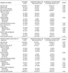
Baseline Characteristics of the Followed-up Subjects with an Equivocal H. pylori Test Finding
kjg-70-181-i001

Values are presented as mean±standard deviation or n (%) unless otherwise indicated.

H. pylori, Helicobacter pylori.

ap-values show the differences between 58 subjects with seroconversion and 40 subjects without seroconversion. Continuous variables are shown as mean value±standard deviation using the Student's t-test. Categorical variables are shown in frequency (%) using the Chi-square test or Fisher's exact test; b≥15 drinks/week for men, ≥8 drinks/week for women.

Table 2

Initial and Follow-up Test Findings of Subjects according to the Follow-up Serum Anti-H. pylori IgG Test Finding

kjg-70-181-i002

Variables are shown as mean value±standard deviation using the Student's t-test.

H. pylori, Helicobacter pylori; IgG, immunoglobulin G; PG, pepsinogen.

ap-values indicate differences between the subjects with seroconversion and without seroconversion; bp-values indicate differences between the initial and follow-up test findings using the signed-rank test.

Table 3

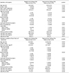
Differences between Subjects with a Negative Test Finding and Those with an Equivocal Test Finding
kjg-70-181-i003

Values are presented as mean±standard deviation or n (%) unless otherwise indicated. Continuous variables are shown as mean value±standard deviation using the Student's t-test. Categorical variables are shown in frequency (%) using the Chi-square test or Fisher's exact test.

H. pylori, Helicobacter pylori; PG, pepsinogen.

Table 4

Findings of Subjects with a History of H. pylori Eradication

kjg-70-181-i004

Upward arrow (↑) indicates that there was a synchronous increase in serum PG I and PG II levels during the follow-up period, whereas downward (↓) indicates vice versa.

H. pylori, Helicobacter pylori; M, male; F, female; PG, pepsinogen.

aSix subjects showed seroconversion at the follow-up test, although the initial serum PG I and PG II levels were low. Most showed decreasing trends of serum PG levels at the follow-up tests, suggesting that seroconversion is related to slow IgG clearance at posteradication period; bThree subjects showed increasing trends of serum PG levels at the follow-up tests, suggesting that there is a reinfection or recrudescence in these subjects; cTwo subjects showed seroreversion despite their high initial serum PG levels. Decreasing trends of serum PG I and PG II levels in these subjects suggest that initial equivocal test findings in these subjects were due to the delayed seroreversion after eradication.

Notes

Financial support This study was supported by the Korean National Research Foundation (NRF 2016R1D1A1B02008937).

Conflict of interest None.

References

1. Sung JK. Diagnosis and treatment of helicobacter pylori infection. Korean J Med. 2015; 89:149–156.
2. Mizuno S, Miki I, Ishida T, et al. Prescreening of a high-risk group for gastric cancer by serologically determined helicobacter pylori infection and atrophic gastritis. Dig Dis Sci. 2010; 55:3132–3137.
3. Choi HS, Lee SY, Kim JH, et al. Combining the serum pepsinogen levels and helicobacter pylori antibody test for predicting the histology of gastric neoplasm. J Dig Dis. 2014; 15:293–298.
4. Samloff IM, Varis K, Ihamaki T, Siurala M, Rotter JI. Relationships among serum pepsinogen I, serum pepsinogen II, and gastric mucosal histology. A study in relatives of patients with pernicious anemia. Gastroenterology. 1982; 83(1 Pt 2):204–209.
5. Samloff IM. Cellular localization of group I pepsinogens in human gastric mucosa by immunofluorescence. Gastroenterology. 1971; 61:185–188.
6. Samloff IM, Liebman WM. Cellular localization of the group II pepsinogens in human stomach and duodenum by immunofluorescence. Gastroenterology. 1973; 65:36–42.
7. Lee SY. Endoscopic gastritis, serum pepsinogen assay, and helicobacter pylori infection. Korean J Intern Med. 2016; 31:835–844.
8. McColl KE. Clinical practice. Helicobacter pylori infection. N Engl J Med. 2010; 362:1597–1604.
9. Ekström AM, Held M, Hansson LE, Engstrand L, Nyrén O. Helicobacter pylori in gastric cancer established by CagA immunoblot as a marker of past infection. Gastroenterology. 2001; 121:784–791.
10. Graham DY, Nurgalieva ZZ, El-Zimaity HM, et al. Noninvasive versus histologic detection of gastric atrophy in a hispanic population in North America. Clin Gastroenterol Hepatol. 2006; 4:306–314.
11. Kokkola A, Rautelin H, Puolakkainen P, et al. Diagnosis of helicobacter pylori infection in patients with atrophic gastritis: comparison of histology, 13C-urea breath test, and serology. Scand J Gastroenterol. 2000; 35:138–141.
12. Groupe d'Etude des Lymphomes Digestifs (GELD) for the Fédération Française de Cancérologie Digestive (FFCD). Lehours P, Ruskone-Fourmestraux A, Lavergne A, Cantet F, Mégraud F. Which test to use to detect helicobacter pylori infection in patients with low-grade gastric mucosa-associated lymphoid tissue lymphoma? Am J Gastroenterol. 2003; 98:291–295.
13. Korstanje A, van Eeden S, Offerhaus JA, et al. Comparison between serology and histology in the diagnosis of advanced gastric body atrophy: a study in a Dutch primary community. J Clin Gastroenterol. 2008; 42:18–22.
14. Testoni PA, Bonassi U, Bagnolo F, Colombo E, Scelsi R. In diffuse atrophic gastritis, routine histology underestimates helicobacter pylori infection. J Clin Gastroenterol. 2002; 35:234–239.
15. Tu H, Sun L, Dong X, et al. Serum anti-helicobacter pylori immunoglobulin G titer correlates with grade of histological gastritis, mucosal bacterial density, and levels of serum biomarkers. Scand J Gastroenterol. 2014; 49:259–266.
16. Chung HA, Lee SY, Moon HW, et al. Does the antibody production ability affect the serum anti-helicobacter pylori IgG titer? World J Gastrointest Pathophysiol. 2016; 7:288–295.
17. Lee SY, Moon HW, Hur M, Yun YM. Validation of western helicobacter pylori IgG antibody assays in Korean adults. J Med Microbiol. 2015; 64(Pt 5):513–518.
18. Kamada T, Hata J, Manabe N, et al. Can dental treatment be the infection route of H. pylori transmission in adults? Three cases of acute gastric mucosal lesions after dental treatment. Dig Endosc. 2007; 19:32–35.
19. Lee JH, Kim N, Chung JI, et al. Long-term follow up of Helicobacter pylori IgG serology after eradication and reinfection rate of H. pylori in South Korea. Helicobacter. 2008; 13:288–294.
20. JPHC Study Group. Tatemichi M, Sasazuki S, Inoue M, Tsugane S. Clinical significance of IgG antibody titer against helicobacter pylori. Helicobacter. 2009; 14:231–236.
21. Lee OJ, Lee EJ, Kim HJ. Correlations among gastric juice pH and ammonia, helicobacter pylori infection and gastric mucosal histology. Korean J Intern Med. 2004; 19:205–212.
22. Huang RG, Xiao HL, Zhou B, et al. Serum pepsinogen levels are correlated with age, sex and the level of helicobacter pylori infection in healthy individuals. Am J Med Sci. 2016; 352:481–486.
23. Lee SP, Lee SY, Kim JH, et al. Correlation between helicobacter pylori infection, IgE hypersensitivity, and allergic disease in Korean adults. Helicobacter. 2015; 20:49–55.
24. Lee SY. Current progress toward eradicating helicobacter pylori in East Asian countries: differences in the 2013 revised guidelines between China, Japan, and South Korea. World J Gastroenterol. 2014; 20:1493–1502.
25. Lee SY. New guidelines for helicobacter pylori treatment: comparisons between Korea and Japan. Korean J Gastroenterol. 2014; 63:151–157.
TOOLS
Similar articles