

 PDF
PDF ePub
ePub Citation
 Print
Print


Abstract
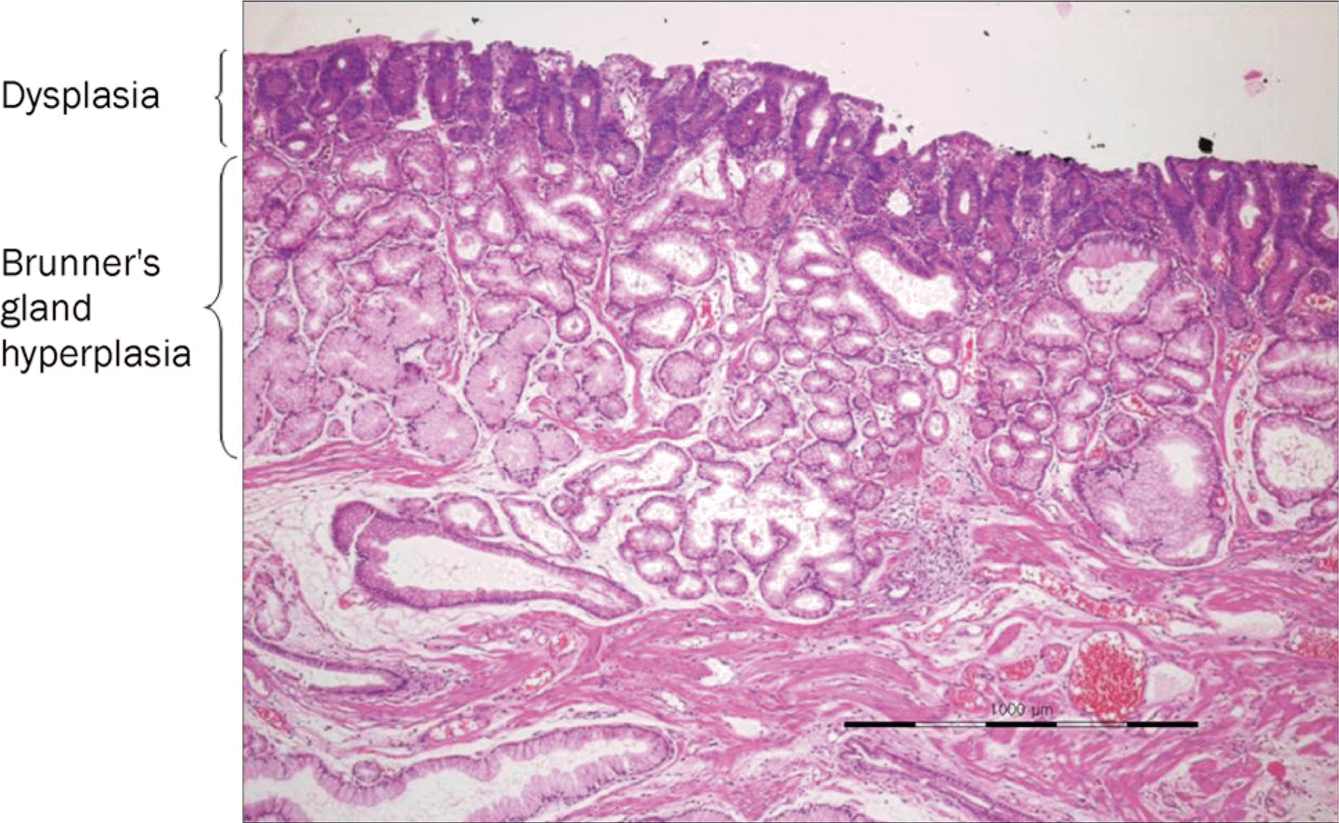
Sporadic non-ampullary duodenal adenoma is uncommon and found incidentally during endoscopic examinations. Brunner's gland hyperplasia is commonly encountered during endoscopic examinations. Adenomas arising from Brunner's gland hyperplasia origi-nate from the glandular cells, and the surface epithelia are usually intact. Little has been reported on adenomas originating from the surface epithelium that overrides Brunner's gland hyperplasia. Here, we report a case of a sporadic non-ampullary duodenal adenoma overriding the cystic dilatation of Brunner's gland hyperplasia.
Go to : 
References
1. Jung SH, Chung WC, Kim EJ, et al. Evaluation of non-ampullary duodenal polyps: comparison of non-neoplastic and neoplastic lesions. World J Gastroenterol. 2010; 16:5474–5480.

2. Kim K, Jang SJ, Song HJ, Yu E. Clinicopathologic characteristics and mucin expression in brunner's gland proliferating lesions. Dig Dis Sci. 2013; 58:194–201.
3. Okada K, Fujisaki J, Kasuga A, et al. Sporadic nonampullary duodenal adenoma in the natural history of duodenal cancer: a study of follow-up surveillance. Am J Gastroenterol. 2011; 106:357–364.
4. Alexander S, Bourke MJ, Williams SJ, Bailey A, Co J. EMR of large, sessile, sporadic nonampullary duodenal adenomas: technical aspects and long-term outcome (with videos). Gastrointest Endosc. 2009; 69:66–73.
5. Honda T, Yamamoto H, Osawa H, et al. Endoscopic submucosal dissection for superficial duodenal neoplasms. Dig Endosc. 2009; 21:270–274.
6. Lépilliez V, Chemaly M, Ponchon T, Napoleon B, Saurin JC. Endoscopic resection of sporadic duodenal adenomas: an efficient technique with a substantial risk of delayed bleeding. Endoscopy. 2008; 40:806–810.
7. Peetz ME, Moseley HS. Brunner's glands hyperplasia. Am Surg. 1989; 55:474–477.
8. Lu L, Li R, Zhang G, Zhao Z, Fu W, Li W. Brunner's gland adenoma of duodenum: report of two cases. Int J Clin Exp Pathol. 2015; 8:7565–7569. eCollection 2015.
Go to :
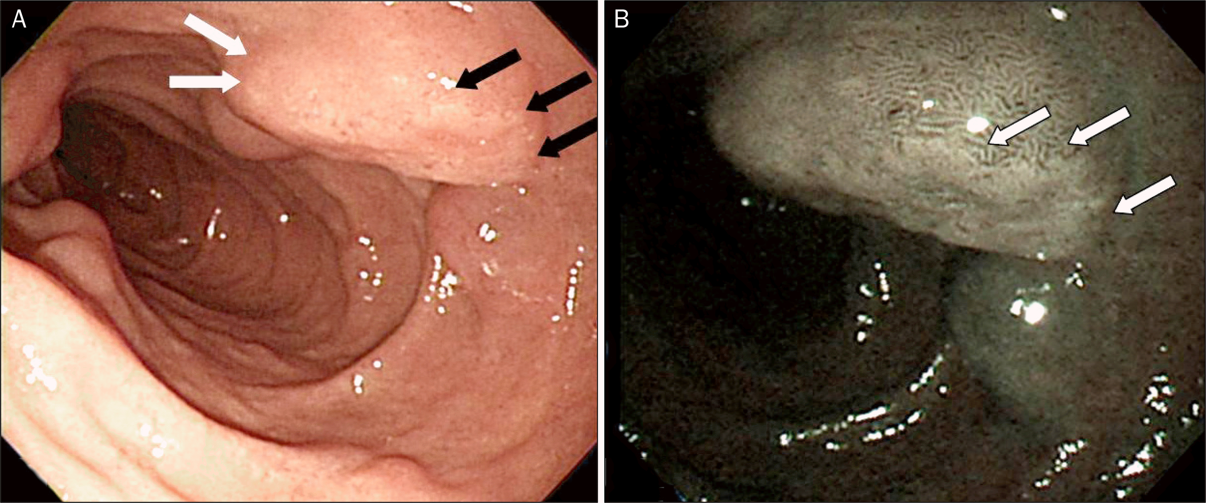 | Fig. 1.Duodenoscopy of the lesion. (A) A nodular lesion (black arrows) is overriding a cystic lesion (white arrows). (B) Narrow band image shows more discrete lesion with different pit patterns (white arrows) compared with background cystic lesions. |
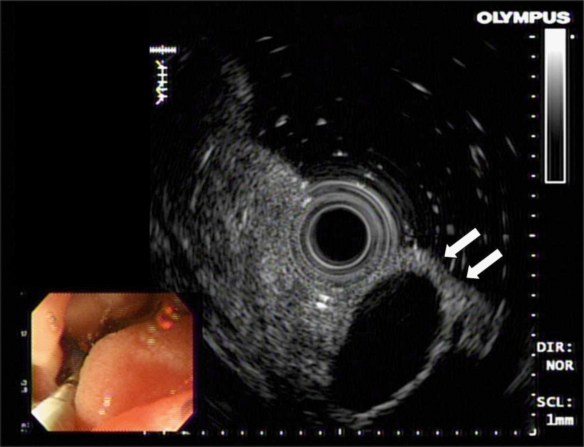 | Fig. 2.Endoscopic ultrasound of the lesion. A hypoechoic homogenous lesion is located at the submucosal layer (white arrows). |

 XML Download