

 PDF
PDF ePub
ePub Citation
 Print
Print


Figures and Tables
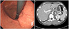
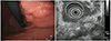 | Fig. 1Initial endoscopy and endosonography. (A) Endoscopy shows a subepithelial lesion with a small, deep ulceration in its center at the posterior wall of the upper body of the stomach. (B) Endosonography reveals that the subepithelial lesion is connected to enlarged perigastric lymph nodes with central hyperechoic area.
|
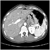 | Fig. 2Initial abdominal computed tomography. Enlargement of perigastric lymph nodes with center hypodense area are observed and these lymph nodes are connected to the gastric wall.
|
References
1. Akgun Y. Intestinal and peritoneal tuberculosis: changing trends over 10 years and a review of 80 patients. Can J Surg. 2005; 48:131–136.
2. Kruijshaar ME, Abubakar I. Increase in extrapulmonary tuberculosis in England and Wales 1999-2006. Thorax. 2009; 64:1090–1095.
3. Horvath KD, Whelan RL. Intestinal tuberculosis: return of an old disease. Am J Gastroenterol. 1998; 93:692–696.
4. Sharma MP, Bhatia V. Abdominal tuberculosis. Indian J Med Res. 2004; 120:305–315.
5. Bandyopadhyay SK, Bandyopadhyay R, Chatterjee U. Isolated gastric tuberculosis presenting as haematemesis. J Postgrad Med. 2002; 48:72–73.
6. Chung JS, Cho YB, Heo WG, Jo DH, Jeong YH, Seo GS. Asymptomatic synchronous tuberculosis involving stomach and small bowel in immunocompetent patient. Korean J Gastroenterol. 2015; 66:345–349.
7. Talukdar R, Khanna S, Saikia N, Vij JC. Gastric tuberculosis presenting as linitis plastica: a case report and review of the literature. Eur J Gastroenterol Hepatol. 2006; 18:299–303.
8. Kim JH, Jeon YC, Kim TY, et al. A case of synchronous intestinal tuberculosis involving the stomach and colon. Korean J Gastroenterol. 2008; 52:320–324.
9. Quantrill SJ, Archer GJ, Hale RJ. Gastric tuberculosis presenting with massive hematemesis in association with acute myeloid leukemia. Am J Gastroenterol. 1996; 91:1259–1260.
 XML Download