

 PDF
PDF ePub
ePub Citation
 Print
Print


INTRODUCTION
Small bowel obstruction is a clinical condition often caused by postoperative adhesion, volvulus, intussusceptions, and hernia. Small bowel obstruction due to bezoars is clinically uncommon, accounting for approximately 2-4% of all obstructions.123 Bezoars are solidified substances formed by a mixture of indigestible exogenous substances with other substances in the gastrointestinal tract. Bezoars are usually formed in the stomach and passed down into the small bowel, resulting in small bowel obstruction.23 Clinically, bezoar-related small bowel obstructions are not easily distinguishable from obstructions caused by other factors.2 Patients with small bowel bezoars usually remain asymptomatic for many years and develop symptoms insidiously. Although computed tomography (CT) is a useful method in diagnosing the cause of small bowel obstruction,4 small bowel obstruction caused by bezoars are often not detected by an abdominal CT examination.
In this report, we present a case of a 74-year-old man with small bowel obstruction caused by Aloe vera bezoars, which were undetected by an abdominal CT. Bezoar should be included in the differential diagnosis of small bowel obstructions when the cause of small bowel obstruction is unclear.
CASE REPORT
A 74-year-old man with intermittent abdominal pain and frequent vomiting visited the emergency room. He previously visited the emergency room of another university hospital, where he was diagnosed with enteritis due to the lack specific findings regarding the small bowel ileus. His abdominal pain was located specifically at the epigastric area, and he experienced six episodes of vomiting. Ten days before admission to our hospital, he experienced intermittent abdominal pain; however, vomiting occurred one day before admission. He denied any weight loss or fever at the time of admission.
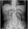
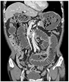
He had past medical history of diabetes mellitus for 11 years. The patient's vital signs on admission were normal. His physical examination revealed distension and tenderness in the epigastric abdomen without rebound tenderness. His bowel sounds were exaggerated. His laboratory data on admission were as follows: white blood cell count, 5,800/mm3; hemoglobin, 13.6 g/dL; platelets, 204,000/mm3; and C-reactive protein levels, 7.2 mg/dL. Mild azotemia was attributed to dehydration and was easily rectified by rehydration. Other findings, including a chemical battery and electrolytes, were unremarkable. A simple abdominal radiograph revealed small bowel ileus with multiple air-fluid levels; however, no free air density was noted (Fig. 1). CT scan of the abdomen and pelvis revealed edematous wall thickening and dilatation of the small bowel without any definite cause of obstruction, such as abnormal mass (Fig. 2).
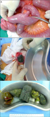
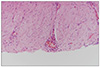
The patient was provided conservative therapy with hydration, parenteral nutrition, and antibiotics to manage the most probable diagnosis, which was partial small bowel obstruction or enteritis of unknown cause. He experienced fart, and improved abdominal pain and vomiting with fasting and regular sips of water for three days. On the fourth day, however, his abdominal pain aggravated after starting his diet. Therefore, a laparoscopic abdominal exploration was performed to confirm the exact cause of small bowel obstruction. The intraoperative findings revealed small bowel ischemia and obstruction caused by an intraluminal mass located in the ileum, 60 cm proximal to the ileocecal valve. We performed a resection of the ischemic small bowel segment and removed the intraluminal mass. The small bowel loops were dilated and obstructed by multiple greenish masses (Fig. 3). Histopathological examination revealed acute serositis of the small intestine with neutrophils, which extended into the intestinal wall, and no other abnormalities were observed (Fig. 4). Postoperatively, we noted that the patient had consumed excessive amounts of Aloe vera, for 10 days, as a natural medicine for dyspepsia. We found round or ovoid intraluminal masses containing mottled air density in the stomach and small bowel via retrospective review of the CT scan of the abdomen and pelvis. The patient recovered without any complications and discharged on the 13th postoperative day.
DISCUSSION
Bezoars are a concretion of indigestible material found in the gastrointestinal tract. Bezoars can be classified into four different groups, according to the component sources: phytobezoars, trichobezoars, pharmacobezoars, and lactobezoars.5 Among these groups, phytobezoars are the most common and are composed of poorly digested fruit and vegetable fibers.6 The occurrence of bezoars is related to diet habits, like ingestion of high-fiber foods, poor mastication, previous gastric surgery, diabetic gastroparesis, and hypothyroidism.4 In the present case, the formation of phytobezoar was related to the ingestion of Aloe vera and diabetic gastroparesis. Aloe vera is rich in fiber, lignins, and tannic acid.7 Tannic acid, when consumed in high amounts, is transformed from a monomeric form into a tannincellulose-protein complex via the actions of the gastric acid. This complex forms a concrete-like viscous mass, incarcerating the insides of the gastrointestinal lumen and ultimately causing obstruction.389 Previously, and to the best of our knowledge, only one case of gastric bezoar by Aloe vera has been reported in the literature.10 However, no case of small bowel obstruction by Aloe vera bezoars has been reported to date.
Clinical manifestation of small bowel bezoars may vary in accordance with different bezoar sizes, obstruction sites, and degrees.11 The most common symptom of bezoar-induced small bowel obstruction is abdominal pain (96-100%), followed by nausea and vomiting;12 similar symptoms were observed in the present case. Bezoars are usually formed in the stomach and passed down into the small bowel, which may lead to small bowel obstruction.13 Small bowel obstruction usually occurs at the narrow part of the small intestine, in particular, the distal end of the ileum following the jejunum. Most bezoars in the small bowel are found 50-70 cm proximal to the ileocecal valve,14 which was consistent with the findings in our case report. Moreover, bezoars may be detected at the anastomosis sites in patients with previous history of gastrointestinal surgery.
Plain abdominal radiography, barium studies, abdominal ultrasound, and abdominal CT scan are used to identify the causes of small bowel obstruction. Among these modalities, abdominal CT is the most useful method in diagnosing small bowel obstructions caused by bezoar, with a diagnostic accuracy of about 73-95%.3 The most common CT finding of bezoars includes a round or ovoid intraluminal mass that contains mottled gas at the obstructed site.115 However, abdominal CT may fail to detect small bowel bezoars, as it can be interpreted as normal intraluminal food materials. To properly manage small bowel obstruction caused by bezoars, early surgical treatment is essential,3 and delayed treatment may significantly increase complications and mortality.10 In accordance with a previous report,16 enterotomy was performed to remove the bezoar in the present case since manual fragmentation was not possible. Segmental bowel resection and anastomosis may be required for ischemia or gangrene-related complications,17 similar to the present case. Morbidity and mortality rates of the surgical approach for small bowel bezoars is 22-32.14% and 2-14.28%, respectively.310
In conclusion, small bowel obstruction caused by Aloe vera bezoars may not be detected by an abdominal CT. Bezoars should be included in the differential diagnosis of small bowel obstruction in patients with predisposing factors, such as excessive consumption of high-fiber foods and diabetes.
 XML Download