Abstract
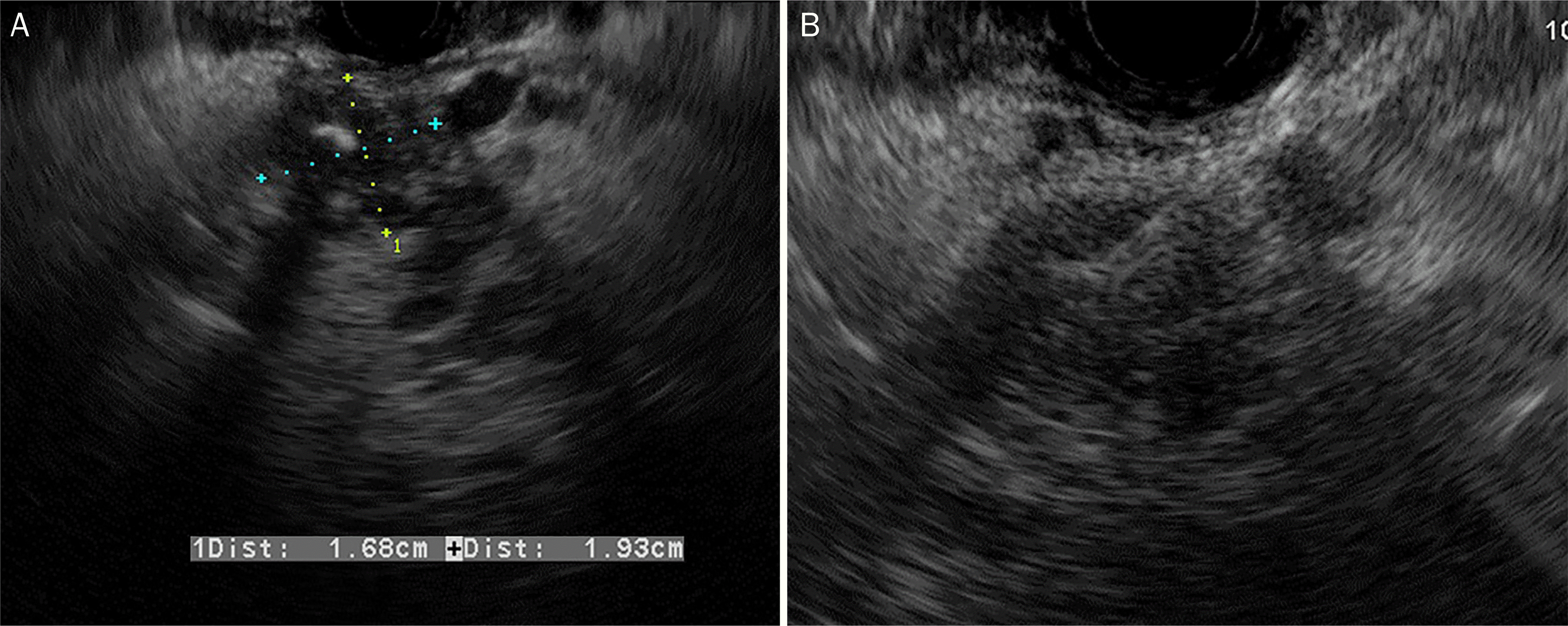
Actinomycosis is a slowly progressive, chronic infectious disease. It is caused by the genus Actinomyces, which are gram-positive anae-robic bacteria. It presents as a mass-like lesion, composed of bacterial nidus and characteristic granulomatous inflammatory fibrosis. As such, it has frequently been mistaken for a malignancy. Surgical resection is a common procedure in these patients prior to a definite diagnosis. Although actinomycosis can occur in a variety of regions, including oral-cervicofacial, thoracic, and abdominopelvic cavities, the involvement of the pancreas is very rare. We report a case of a 44-year-old male with a symptomatic actinomycosis caused by a mass in the tail of the pancreas. The diagnosis was made using an endoscopic ultrasound-guided fine needle aspiration biopsy without surgical resection. After the treatment with antibiotics, the pancreatic mass was confirmed to be resolved on the follow-up computed tomography.
Go to : 
References
3. Kim MC, Lee H, Park J, et al. A solitary pancreatic actinomycosis mimicking pancreatic cancer. Korean J Pancreas Biliary Tract. 2015; 20:130–135.

4. Lee JH, Lee KG, Oh YH, Park HK, Lee KS. Actinomycosis of the pancreas: a case report and review of the literature. Hepatogastroenterology. 2010; 57:358–361.
5. Maestro S, Trujillo R, Geneux K, et al. Pancreatic actinomycosis presenting as pancreatic mass and diagnosed with endoscopic ultrasound fine needle aspiration (EUS-FNA). Endoscopy. 2013; 45(Suppl 2 UCTN):E276–E277.

6. Piper MH, Schaberg DR, Ross JM, Shartsis JM, Orzechowski RW. Endoscopic detection and therapy of colonic actinomycosis. Am J Gastroenterol. 1992; 87:1040–1042.
7. Harsch IA, Benninger J, Niedobitek G, et al. Abdominal actinomycosis: complication of endoscopic stenting in chronic pancreatitis? Endoscopy. 2001; 33:1065–1069.

8. Kuesters S, Timme S, Keck T. Uncommon cause of an inflammatory pancreatic head tumor. Diagnosis: purulent actinomycosis and incidental T1-carcinoid of the pancreatic head. Gastroenterology. 2011; 141:e9–e10.
9. Somsouk M, Shergill AK, Grenert JP, Harris H, Cello JP, Shah JN. Actinomycosis mimicking a pancreatic head neoplasm diagnosed by EUS-guided FNA. Gastrointest Endosc. 2008; 68:186–187.

10. Hsu JT, Lo HC, Jan YY, Chen HM. Actinomycosis mimicking recurrent carcinoma after whipple's operation. World J Gastroenterol. 2005; 11:1722–1724.

11. Harris LA, DeCosse JJ, Dannenberg A. Abdominal actinomycosis: evaluation by computed tomography. Am J Gastroenterol. 1989; 84:198–200.
12. Lee IJ, Ha HK, Park CM, et al. Abdominopelvic actinomycosis involving the gastrointestinal tract: CT features. Radiology. 2001; 220:76–80.

13. Cintron JR, Del Pino A, Duarte B, Wood D. Abdominal actinomycosis. Dis Colon Rectum. 1996; 39:105–108.

14. Ferrari TC, Couto CA, Murta-Oliveira C, Conceição SA, Silva RG. Actinomycosis of the colon: a rare form of presentation. Scand J Gastroenterol. 2000; 35:108–109.
Go to : 
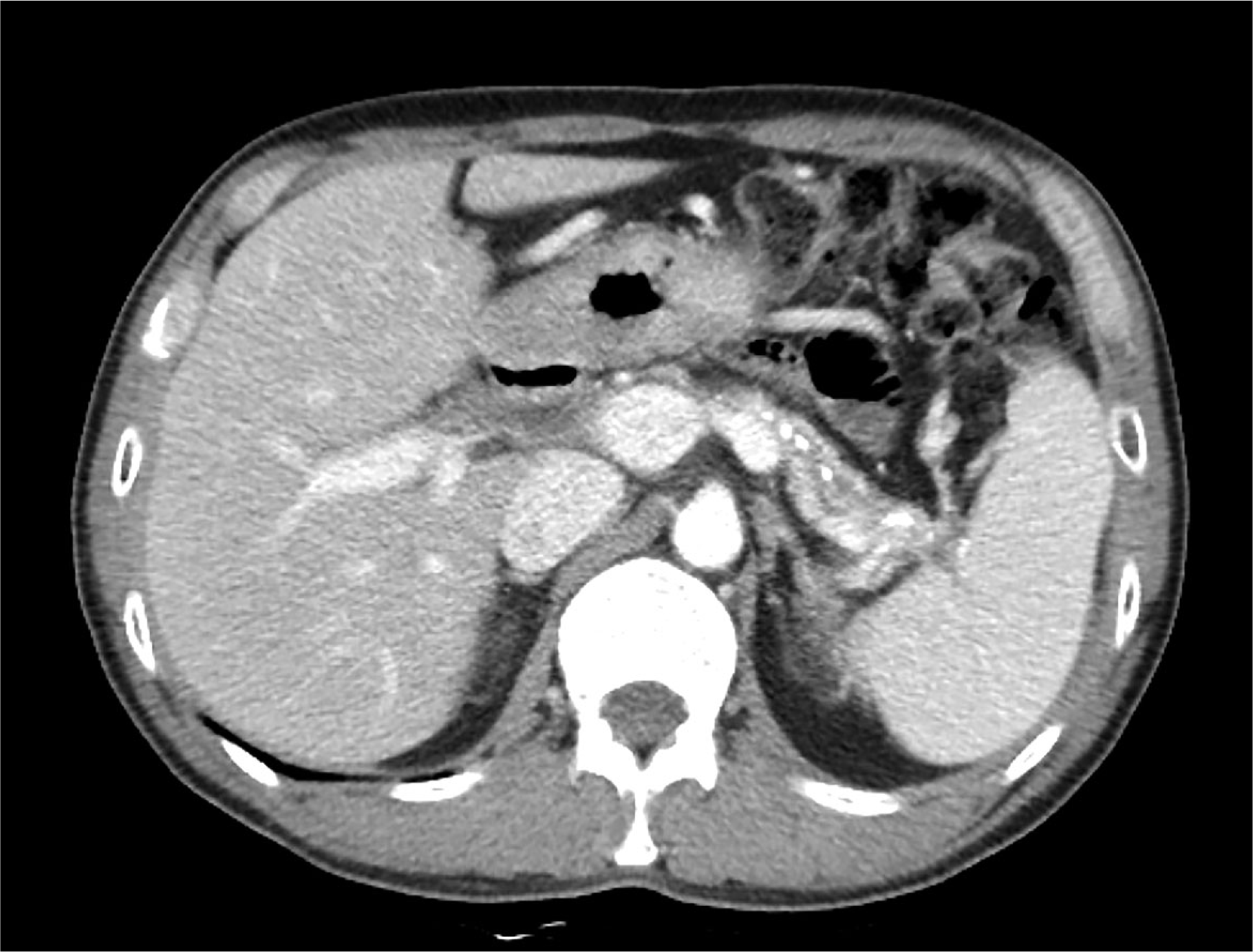
 | Fig. 1.Computed tomography scan images. (A) Multiple calcifications and stones (black arrows) were also noted in the head of the pancreas. (B) Contrast-enhanced axial image (portal phase) revealed diffuse parenchymal swelling with multiple calcification (black arrows) at the body of the pancreas. (C) Contrast-enhanced coronal image (portal phase) showed heterogeneous enhancement of the pancreas parenchyma and peripancreatic strands (white arrows). (D) Multifocal uneven pancreatic duct dilation (white asterisk) was also observed. |
 | Fig. 2.Endoscopic ultrasonography (EUS) images. (A) A 19-mm sized, ill-defined, hypoechoic and heterogenous mass with central calcification was identified at the pancreatic tail. (B) EUS-guided fine needle aspiration biopsy using a 20-gauge needle was performed. |
 | Fig. 3.Microscopic findings for the specimen. The specimen revealed sulfur granules, consisting of a conglomeration of filamentous bacteria, which was shown at the surrounding tissue with dense inflammatory infiltration by lymphocytes, neutrophils, and foamy macrophages (A, hematoxylin and eosin stain, ×100; B, hematoxylin and eosin stain, ×400). |


 PDF
PDF ePub
ePub Citation
 Print
Print


 XML Download