Abstract
Multiple endocrine neoplasia type 1 (MEN1) syndrome is a relatively rare disease, characterized by the occurrence of multiple endocrine tumors in the parathyroid and pituitary glands as well as the pancreas. Here, we report a case of MEN1 with neuroendocrine tumors (NETs) in the stomach, duodenum, and pancreas. A 53-year-old man visited our hospital to manage gastric NET. Five years prior to his visit, he had undergone surgery for incidental meningioma. His brother had pancreatic nodules and a history of surgery for adrenal adenoma. His brother's daughter also had pancreatic nodules, but had not undergone surgery. The lesion was treated by endoscopic submucosal dissection and diagnosed as a grade 1 NET. Another small NET was detected in the second duodenal portion, resected by endoscopic submucosal dissection, which was also diagnosed as a grade 1 NET. During evaluation, three nodules were detected in the pancreas, and no evidence of pituitary, parathyroid tumors, or metastasis was observed. After surgery, the pancreatic lesions were diagnosed as NETs, with the same immunohistochemical patterns as those of the stomach and duodenum. Genetic testing was performed, and a heterozygous mutation was detected in the MEN1 gene, which is located on 11q13.
Go to : 
References
1. Kaltsas GA, Besser GM, Grossman AB. The diagnosis and medical management of advanced neuroendocrine tumors. Endocr Rev. 2004; 25:458–511.

2. Marx S, Spiegel AM, Skarulis MC, Doppman JL, Collins FS, Liotta LA. Multiple endocrine neoplasia type 1: clinical and genetic topics. Ann Intern Med. 1998; 129:484–494.

3. Thakker RV, Newey PJ, Walls GV, et al. Clinical practice guidelines for multiple endocrine neoplasia type 1 (MEN1). J Clin Endocrinol Metab. 2012; 97:2990–3011.

4. Brandi ML, Gagel RF, Angeli A, et al. Guidelines for diagnosis and therapy of MEN type 1 and type 2. J Clin Endocrinol Metab. 2001; 86:5658–5671.
5. Larsson C, Skogseid B, Oberg K, Nakamura Y, Nordenskjold M. Multiple endocrine neoplasia type 1 gene maps to chromosome 11 and is lost in insulinoma. Nature. 1988; 332:85–87.

6. Lips CJ, Dreijerink KM, Höppener JW. Variable clinical expression in patients with a germline MEN1 disease gene mutation: clues to a genotype-phenotype correlation. Clinics (Sao Paulo). 2012; 67(Suppl 1):49–56.

7. Lemos MC, Thakker RV. Multiple endocrine neoplasia type 1 (MEN1): analysis of 1336 mutations reported in the first decade following identification of the gene. Hum Mutat. 2008; 29:22–32.

8. Agarwal SK, Kester MB, Debelenko LV, et al. Germline mutations of the MEN1 gene in familial multiple endocrine neoplasia type 1 and related states. Hum Mol Genet. 1997; 6:1169–1175.

9. Schaaf L, Pickel J, Zinner K, et al. Developing effective screening strategies in multiple endocrine neoplasia type 1 (MEN 1) on the basis of clinical and sequencing data of German patients with MEN 1. Exp Clin Endocrinol Diabetes. 2007; 115:509–517.

10. Pipeleers-Marichal M, Somers G, Willems G, et al. Gastrinomas in the duodenums of patients with multiple endocrine neoplasia type 1 and the Zollinger-Ellison syndrome. N Engl J Med. 1990; 322:723–727.

11. Carriaga MT, Henson DE. Liver, gallbladder, extrahepatic bile ducts, and pancreas. Cancer. 1995; 75(1 Suppl):171–190.

12. Triponez F, Dosseh D, Goudet P, et al. Epidemiology data on 108 MEN 1 patients from the GTE with isolated nonfunctioning tumors of the pancreas. Ann Surg. 2006; 243:265–272.

13. Halfdanarson TR, Rabe KG, Rubin J, Petersen GM. Pancreatic neuroendocrine tumors (PNETs): incidence, prognosis and recent trend toward improved survival. Ann Oncol. 2008; 19:1727–1733.

14. Donegan D, Singh Ospina N, Rodriguez-Gutierrez R, et al. Longterm outcomes in patients with multiple endocrine neoplasia type 1 and pancreaticoduodenal neuroendocrine tumours. Clin Endocrinol (Oxf). 2017; 86:199–206.

15. Soga J. Endocrinocarcinomas (carcinoids and their variants) of the duodenum. An evaluation of 927 cases. J Exp Clin Cancer Res. 2003; 22:349–363.
16. Vernooij MW, Ikram MA, Tanghe HL, et al. Incidental findings on brain MRI in the general population. N Engl J Med. 2007; 357:1821–1828.

17. Asgharian B, Chen YJ, Patronas NJ, et al. Meningiomas may be a component tumor of multiple endocrine neoplasia type 1. Clin Cancer Res. 2004; 10:869–880.

18. Chang JH, Kim SW, Chung WC, et al. Clinical review of gastrointestinal carcinoid tumor and analysis of the factors predicting metastasis. Korean J Gastroenterol. 2007; 50:19–25.
19. Sun JM, Jung HC. Gastrointestinal carcinoid tumor. Korean J Gastroenterol. 2004; 44:59–65.
Go to : 
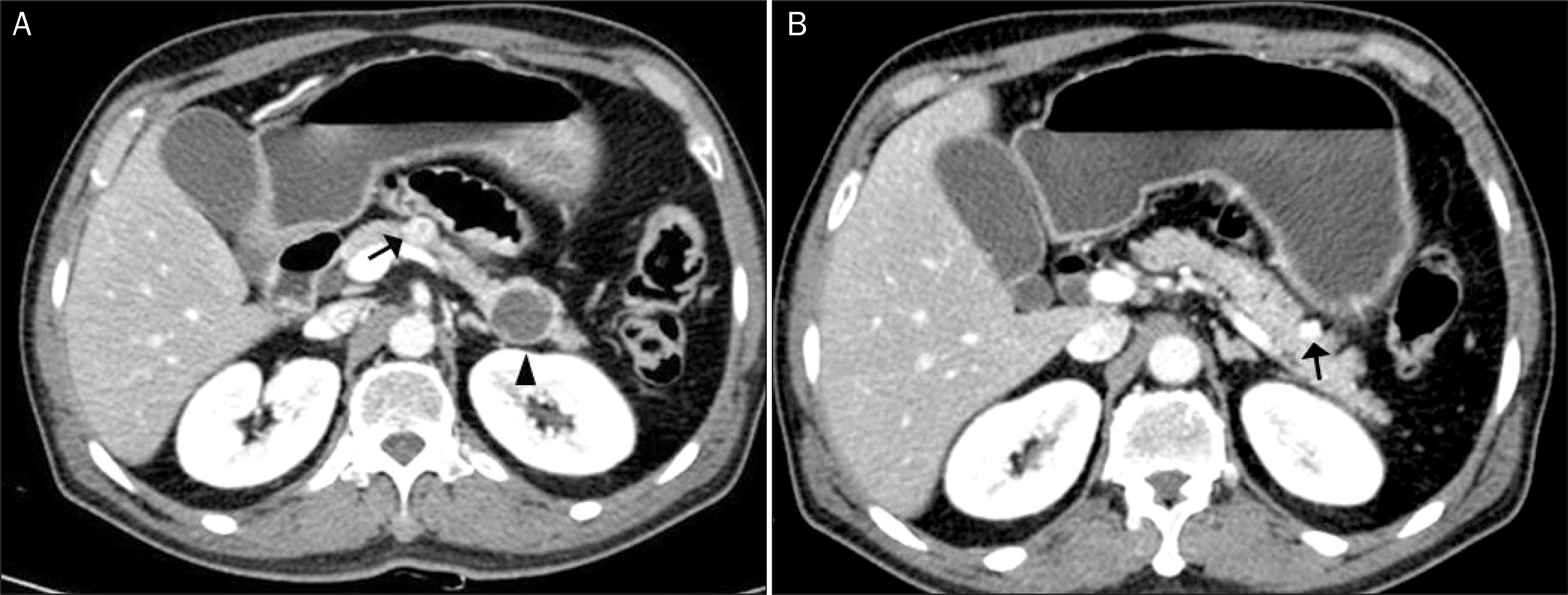
 | Fig. 1.Abdominal computed tomography scan. (A) An arterial enhancing nodule in the pancreatic neck (arrow) and one thick-walled cystic nodule with a mural component and marginal calcification in the tail (arrowhead). (B) Another arterial enhancing nodule in the pancreatic tail (arrow). |
 | Fig. 2.Endoscopic findings for the neuroendocrine tumors in the stomach. (A) A raised nodule with a whitish color and 8×8 mm in the area of the lesser curvature at the lower body of the stomach (arrow). (B) Endoscopic submucosal dissection of the gastric lesion using a soft transparent hood. (C) The completely resected gastric lesion. |
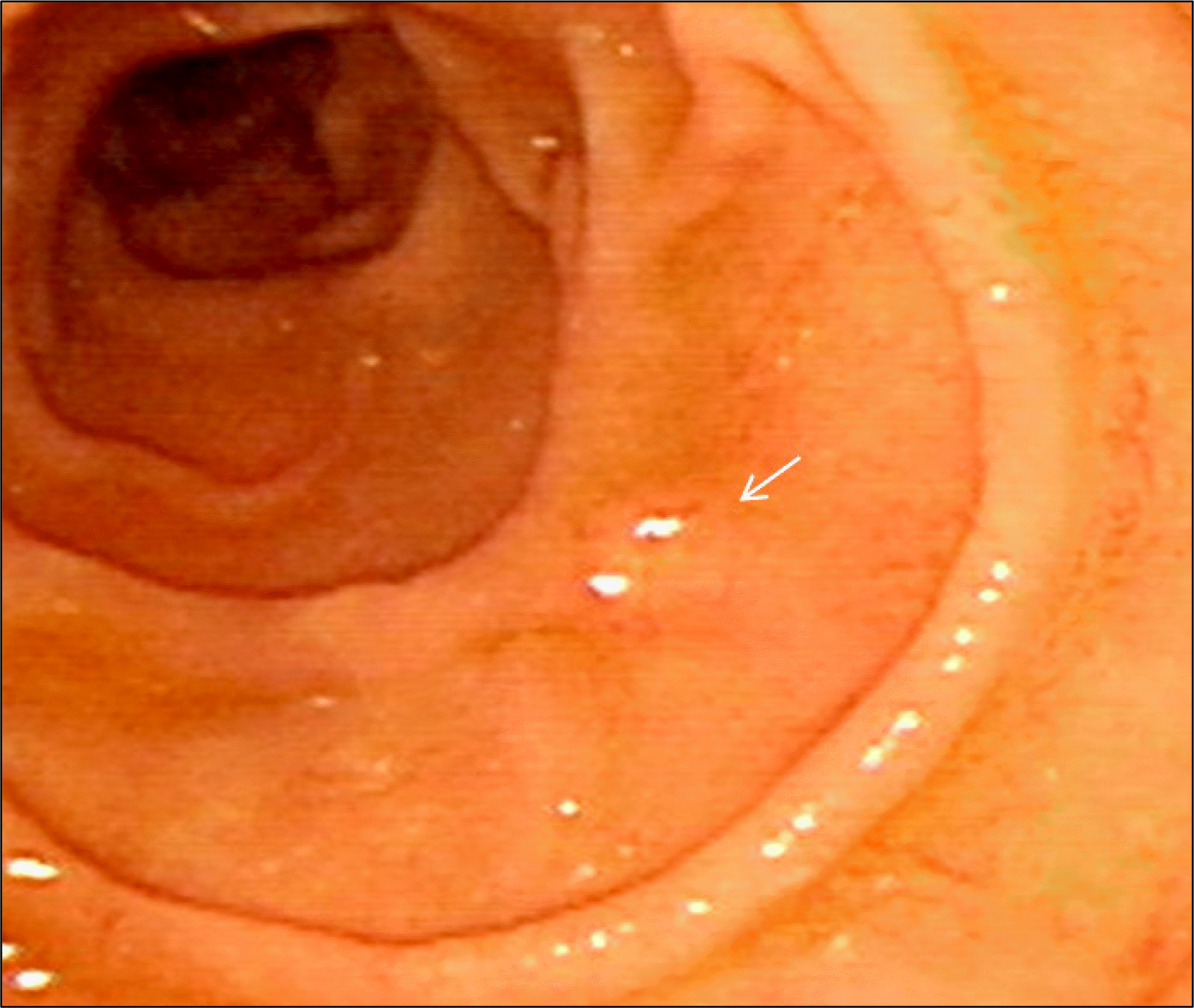
 | Fig. 3.Endoscopic findings for a 3×3 mm yellowish nodule in the second duodenal portion (arrow). |
 | Fig. 4.Pathologic findings after endoscopic submucosal dissection of a gastric neuroendocrine tumor invading the submucosa and mucosa. (A) Tumor cells forming a mostly glandular and trabecular pattern (H&E, ×100). (B) Immunoreactivity for synaptophysin (synaptophysin, ×100). |
 | Fig. 5.Pathologic findings for an endoscopically-resected mucosal specimen containing a duodenal neuroendocrine tumor. (A) Neuroendocrine tumor (arrow) and normal duodenal epithelium (arrowhead) (H&E, ×100). (B) Monotonous proliferation of small round cells with hyperchromatic nuclei and scant cytoplasm forming nests, exhibiting a trabecular growth pattern (H&E, ×100). (C) Immunoreactivity for synaptophysin (synaptophysin, ×100). |


 PDF
PDF ePub
ePub Citation
 Print
Print


 XML Download