

 PDF
PDF ePub
ePub Citation
 Print
Print


Abstract
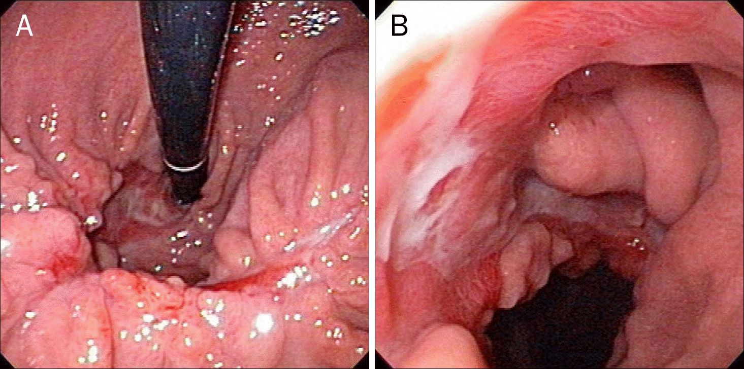
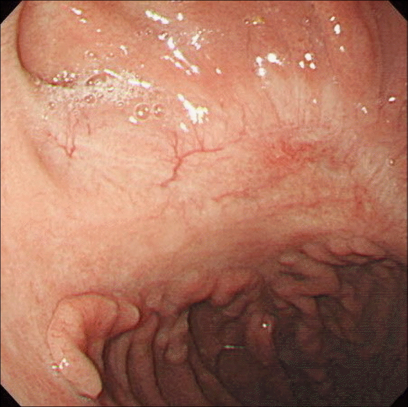
Gastric mucormycosis is a rare and life-threatening fungal disease, caused by fungus in the order Mucorales. While rhino-cerebral and pulmonary forms are common, gastric mucormycosis is an uncommon site for the disease. We diagnosed gastric mucormycosis in a 41-year-old female who had severe multiple trauma, including cardiac rupture, due to a traffic accident. Eighteen days after hospitalization, she passed 800 mL of melena over one day. We performed upper esophagogastroduodenoscopy (EGD) and found a huge gastric ulcer with bleeding. Histopathological examination identified non-septated and right-angled branching fungal hyphae, and we diagnosed gastric mucormycosis. We recommended total gastrectomy to her but she refused the operation, so she was treated with liposomal amphotericin B for 53 days. After two months of treatment with liposomal amphotericin B, we again performed EGD and found a healed gastric ulcer. After four months, with another EGD, we found that the gastric mucormycosis was completely healed.
Go to : 
References
1. Jung JH, Choi HJ, Yoo J, Kang SJ, Lee KY. Emphysematous gastritis associated with invasive gastric mucormycosis: a case report. J Korean Med Sci. 2007; 22:923–927.

2. Camara-Lemarroy CR, González-Moreno EI, Rodríguez-Gutiérrez R, et al. Clinical features and outcome of mucormycosis. Interdiscip Perspect Infect Dis. 2014; 2014:562610.
3. Ha TS, Park CM, Yang JH, et al. Disseminated gastrointestinal mucormycosis in immunocompromised disease. Korean J Crit Care Med. 2015; 30:323–328.
4. Thomson SR, Bade PG, Taams M, Chrystal V. Gastrointestinal mucormycosis. Br J Surg. 1991; 78:952–954.
5. Lee JS, Kim HC, Park SW, et al. A case of isolated pulmonary mucormycosis in an immunocompetent host. Tuberc Respir Dis (Seoul). 2013; 74:269–273.
6. Pahwa M, Pahwa AR, Girotra M, Chawla A. Isolated renal mucormycosis in a healthy immunocompetent patient: atypical presentation and course. Korean J Urol. 2013; 54:641–643.
7. Moon WJ, Kim BJ, Ko YJ, et al. A case of gastric ulcer associated with mucormycosis. Korean J Med. 1999; 56:532–536.
8. Shiva Prasad BN, Shenoy A, Nataraj KS. Primary gastrointestinal mucormycosis in an immunocompetent person. J Postgrad Med. 2008; 54:211–213.
9. Hahn HS, Jung HS, Song SH, et al. A case intestinal mucormycosis healing of ulcer after only amphotericin B treatment. Korean J Gastrointest Endosc. 2002; 25:43–47.
10. Choi WR, Lim CN, Won KH, et al. A case of gastric mucormycosis associated with diabetes mellitus and uremia. Korean J Gastrointest Endosc. 1999; 19:953–958.
11. Kim JS, Ko YW, Jang JH, et al. A case of mucormycosis in a patient with myelodysplastic syndrome and review of the literature in Korea. Korean J Infect Dis. 1999; 31:425–434.
12. Berne JD, Villarreal DH, McGovern TM, Rowe SA, Moore FO, Norwood SH. A fatal case of posttraumatic gastric mucormycosis. J Trauma. 2009; 66:933–935.
13. Johnson CB, Ahmeti M, Tyroch AH, Zuckerman MJ, Hakim MN. Gastric mucormycosis as a cause of life-threatening upper gastrointestinal bleeding in a trauma patient. Am Surg. 2010; 76:E76–E77.
14. Stamm B. Mucormycosis of the stomach in a patient with multiple trauma. Histopathology. 2005; 47:222–223.
15. Lyon DT, Schubert TT, Mantia AG, Kaplan MH. Phycomycosis of the gastrointestinal tract. Am J Gastroenterol. 1979; 72:379–394.
16. Kim J, Lee JH, Byeon JS, Jung HC, Song IS, Kim CY. Gastric mucormycosis in a renal transplant recipient. Korean J Gastrointest Endosc. 1998; 18:230–237.
Go to :
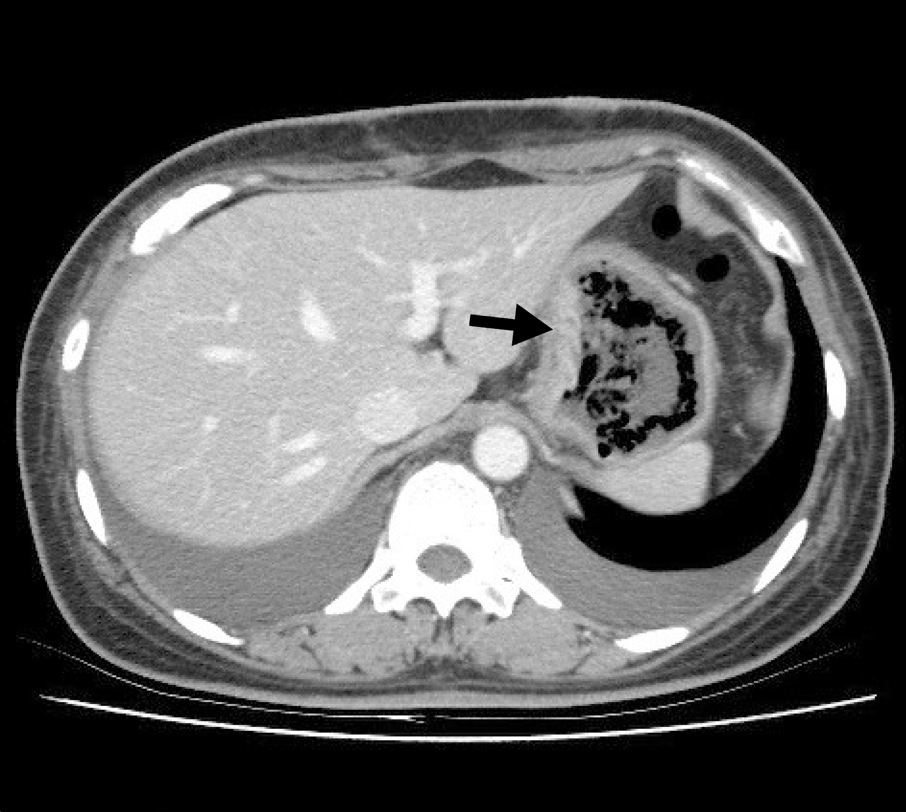 | Fig. 1.Abdominal CT finding. Irregular gastric wall thickening and air-bubble formation at gastric wall (arrow) were seen. Both pleural effusions were also found. |
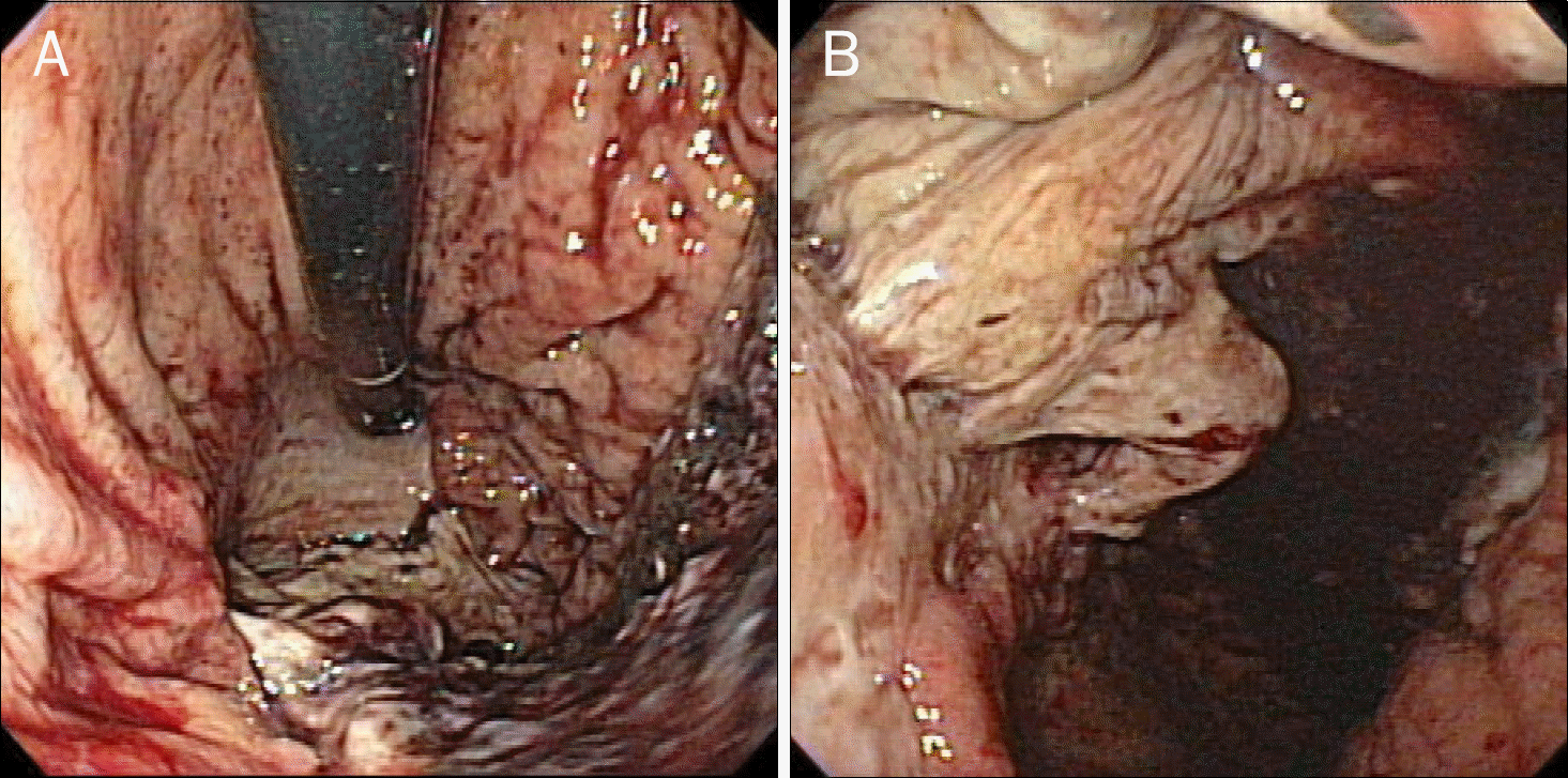 | Fig. 2.Endoscopic findings after melena episode. Huge gastric ulcer with bleeding at great curvature side of gastric body and fundus, covered with yellowish exudate. |
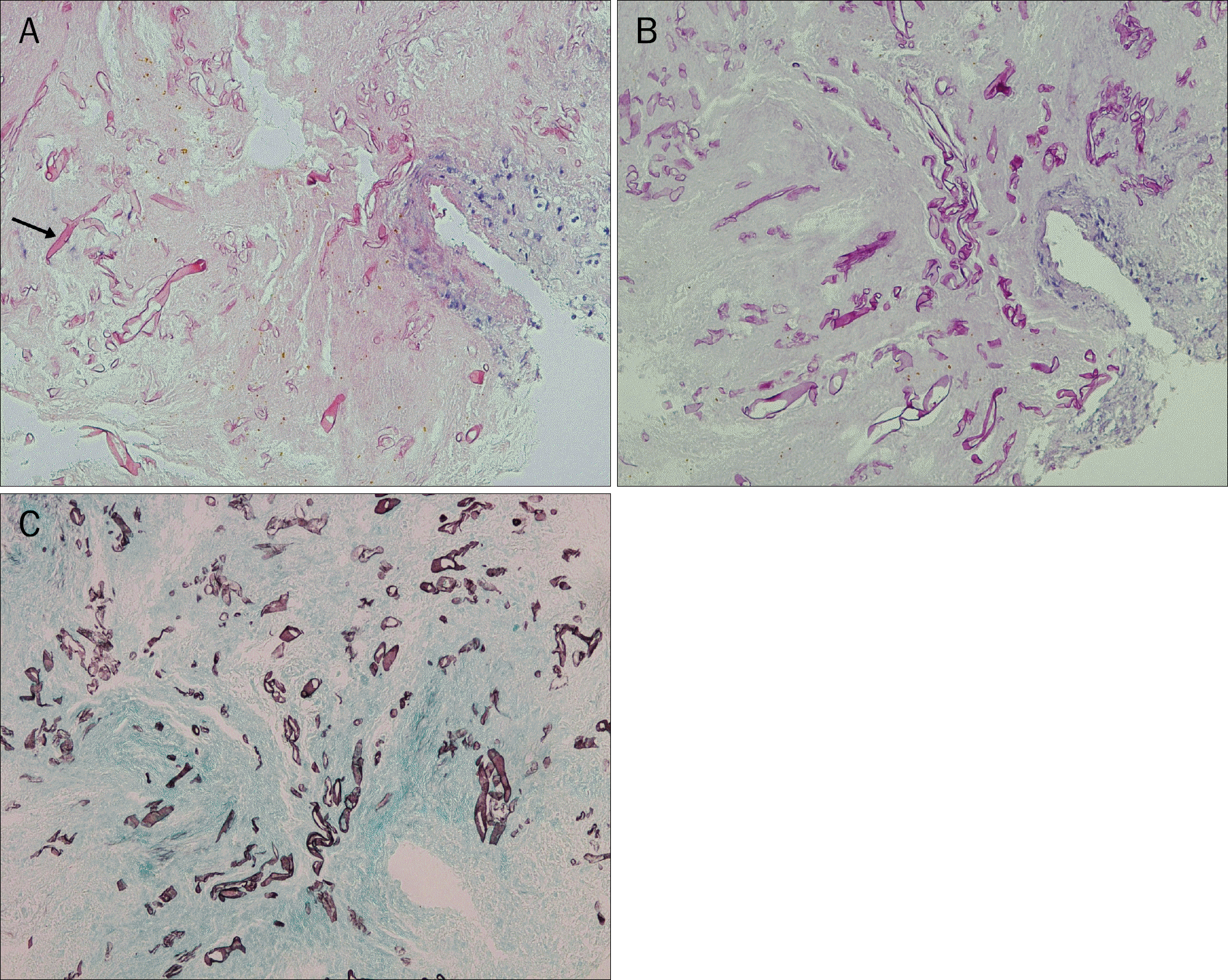 | Fig. 3.Histopathologic findings shown as multiple broad-based, non-septated, right-angle branched fungal hyphae (arrow) with tissue infiltration in H&E (A, ×200), periodic acid-schiff stain (B, ×200), and Gomori methenamine silver stain (C, ×200). |
 XML Download