Abstract
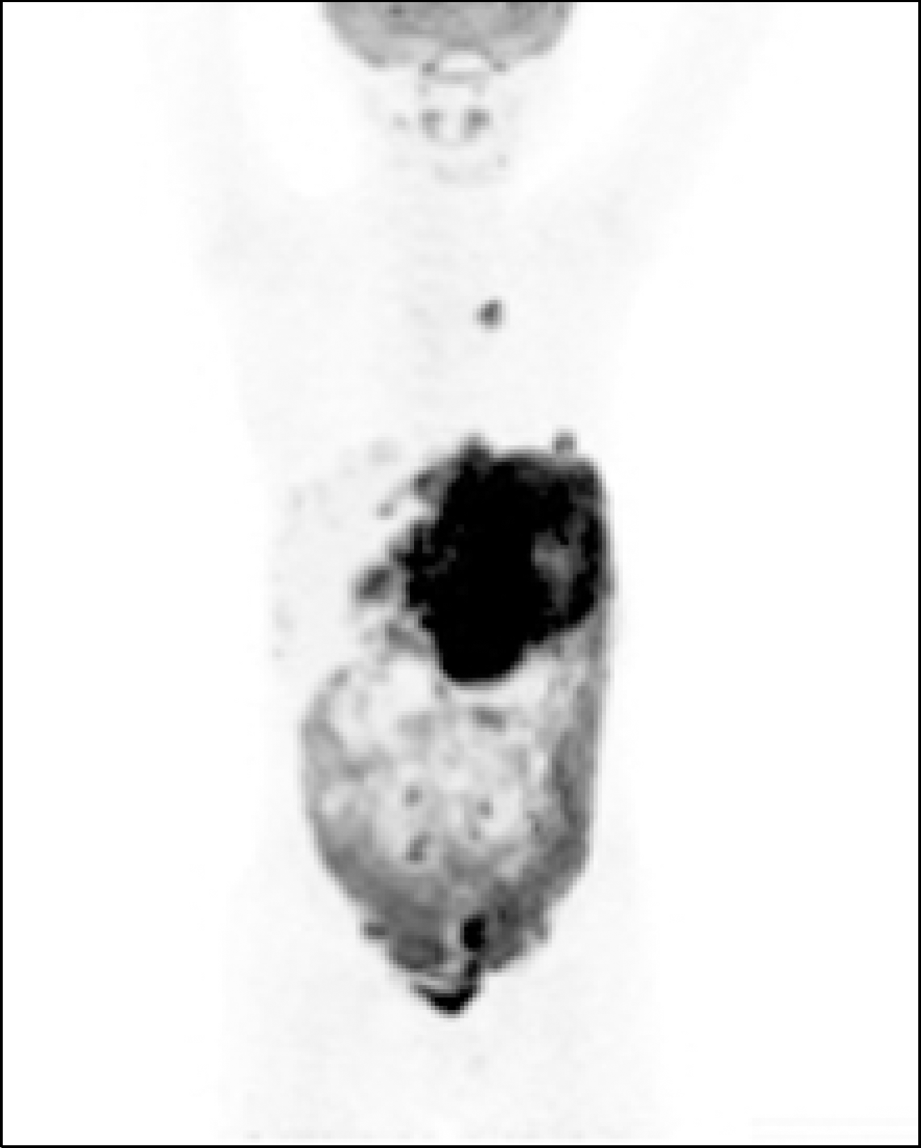
Primary gastric tumors are very rare in children. Burkitt lymphoma is a common type of non-Hodgkin's lymphoma, and gastric Burkitt lymphoma usually occurs in the aged. When involving the gastrointestinal tract, primary gastric Burkitt lymphoma is very rare in younger childhood. Many gastric lymphomas including mucosa-associated lymphoid tissue lymphoma are associated with Helicobacter pylori infection or acute bleeding symptom. We report a seven-year-old boy who presented with only some vomiting and postprandial pain. His upper gastrointestinal endoscopy and biopsy revealed a large primary Burkitt lymphoma with no acute bleeding and no evidence of H. pylori infection. After chemotherapy, he remains in remission.
References
1. Lee SH, Kim HJ, Mun JS, et al. A case of primary hepatic Burkitt's lymphoma. Korean J Gastroenterol. 2008; 51:259–264.
2. Ladd AP, Grosfeld JL. Gastrointestinal tumors in children and adolescents. Semin Pediatr Surg. 2006; 15:37–47.

3. Allen CE, Kelly KM, Bollard CM. Pediatric lymphomas and histio-cytic disorders of childhood. Pediatr Clin North Am. 2015; 62:139–165.

4. Kassira N, Pedroso FE, Cheung MC, Koniaris LG, Sola JE. Primary gastrointestinal tract lymphoma in the pediatric patient: review of 265 patients from the SEER registry. J Pediatr Surg. 2011; 46:1956–1964.

5. Kesik V, Safali M, Citak EC, Kismet E, Koseoglu V. Primary gastric Burkitt lymphoma: a rare cause of intraabdominal mass in childhood. Pediatr Surg Int. 2010; 26:927–929.

6. Pickett LK, Briggs HC. Cancer of the gastrointestinal tract in childhood. Pediatr Clin North Am. 1967; 14:223–234.

7. Ferlay J, Soerjomataram I, Dikshit R, et al. Cancer incidence and mortality worldwide: sources, methods and major patterns in GLOBOCAN 2012. Int J Cancer. 2015; 136:E359–E386.

8. Curtis JL, Burns RC, Wang L, Mahour GH, Ford HR. Primary gastric tumors of infancy and childhood:54-year experience at a single institution. J Pediatr Surg. 2008; 43:1487–1493.
9. Kim HY, Park JH. The role of endoscopy for tumorous conditions of the upper gastrointestinal tract in children. Korean J Pediatr Gastroenterol Nutr. 2005; 8:31–40.

10. Kang G, Park YS, Jung ES, et al. Gastrointestinal stromal tumors in children and young adults: a clinicopathologic and molecular genetic study of 22 Korean cases. APMIS. 2013; 121:938–944.

11. Khurshed A, Ahmed R, Bhurgri Y. Primary gastrointestinal malignancies in childhood and adolescence–an Asian perspective. Asian Pac J Cancer Prev. 2007; 8:613–617.
12. Kamona AA, El-Khatib MA, Swaidan MY, et al. Pediatric Burkitt's lymphoma: CT findings. Abdom Imaging. 2007; 32:381–386.

13. Howell JM, Auer-Grzesiak I, Zhang J, Andrews CN, Stewart D, Urbanski SJ. Increasing incidence rates, distribution and histological characteristics of primary gastrointestinal non-Hodgkin lymphoma in a North American population. Can J Gastroenterol. 2012; 26:452–456.

14. Bautista-Quach MA, Ake CD, Chen M, Wang J. Gastrointestinal lymphomas: morphology, immunophenotype and molecular features. J Gastrointest Oncol. 2012; 3:209–225.
15. Perkins AS, Friedberg JW. Burkitt lymphoma in adults. Hematology Am Soc Hematol Educ Program. 2008; 2008:341–348.

16. Olaniyi JA. Burkitt lymphoma: a review. Sci Rep. 2012; 1:337. 1–4.
17. Baumgaertner I, Copie-Bergman C, Levy M, et al. Complete remission of gastric Burkitt's lymphoma after eradication of Helicobacter pylori. World J Gastroenterol. 2009; 15:5746–5750.
Fig. 1.
Simple abdominal X-ray shows mass-like lesion in the epigastric and in the left infra-phrenic area.
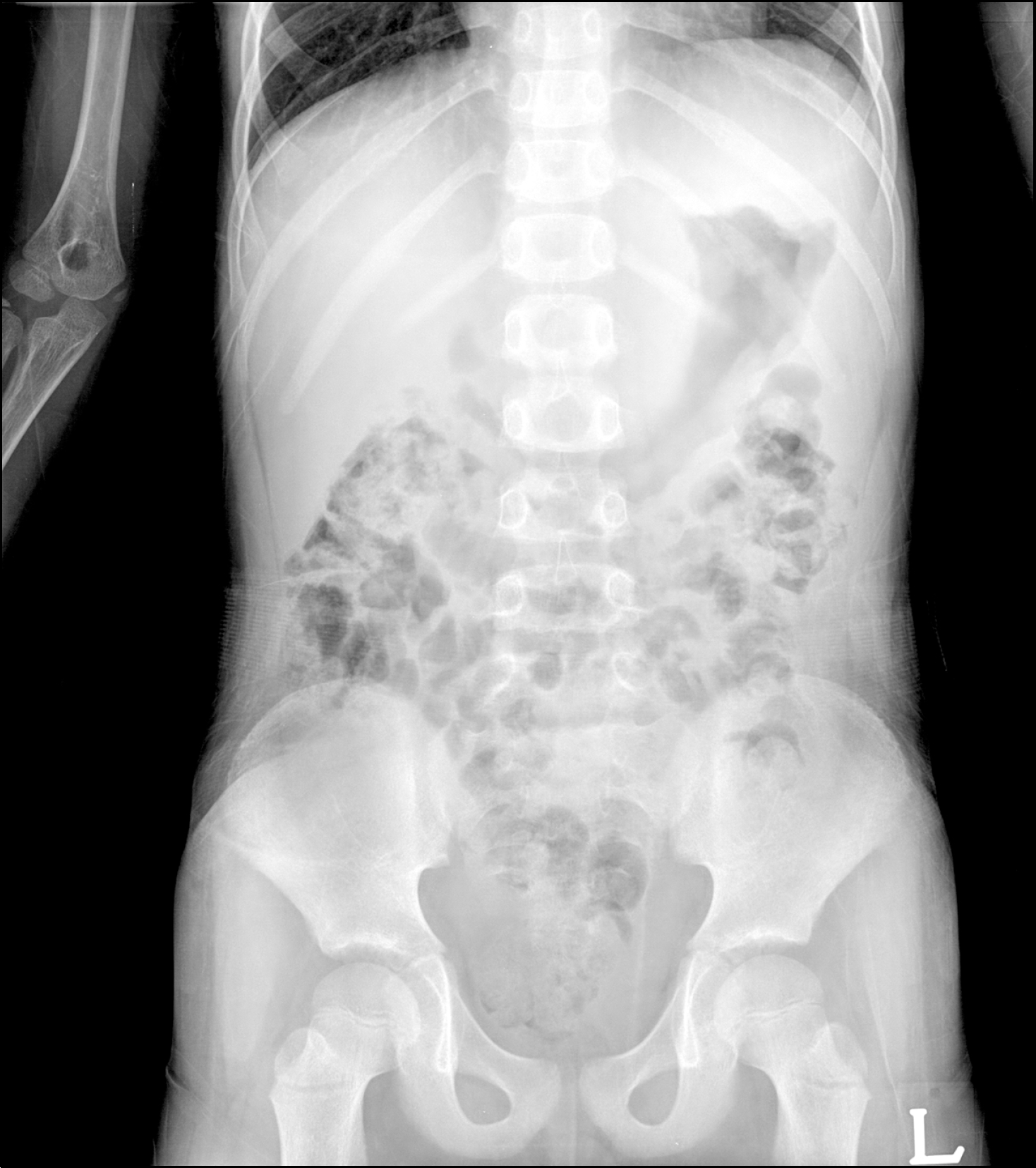
Fig. 2.
Contrast-enhanced coronal CT reveals diffuse thickening of the gastric wall with heterogeneous enhancement.
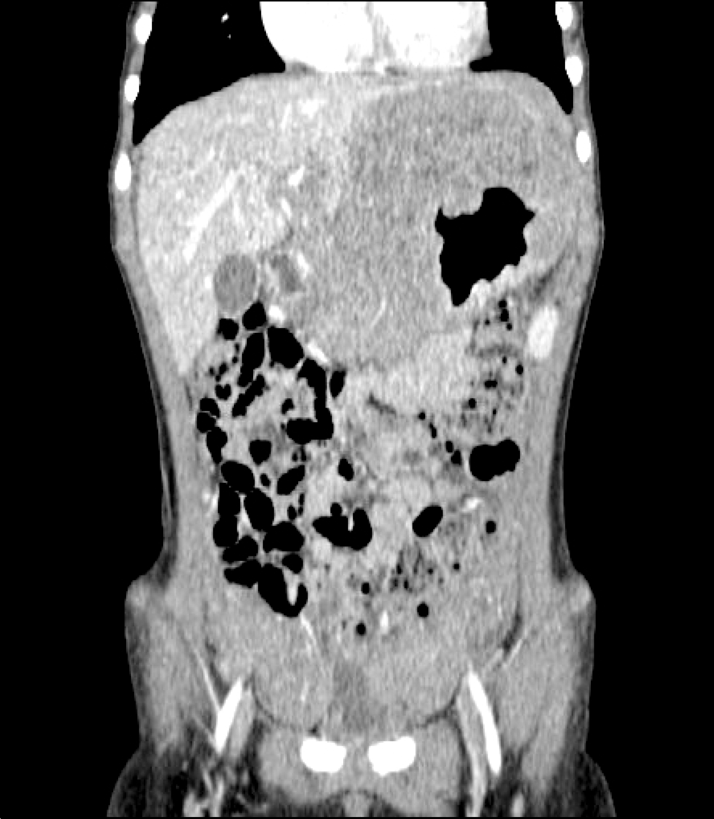
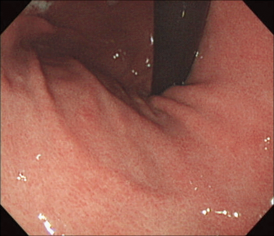
Fig. 3.
Gastroscopic finding. A huge well-demarcated ulcerative lesion with neighboring mucosal elevation and irregular margin is seen at the cardia.
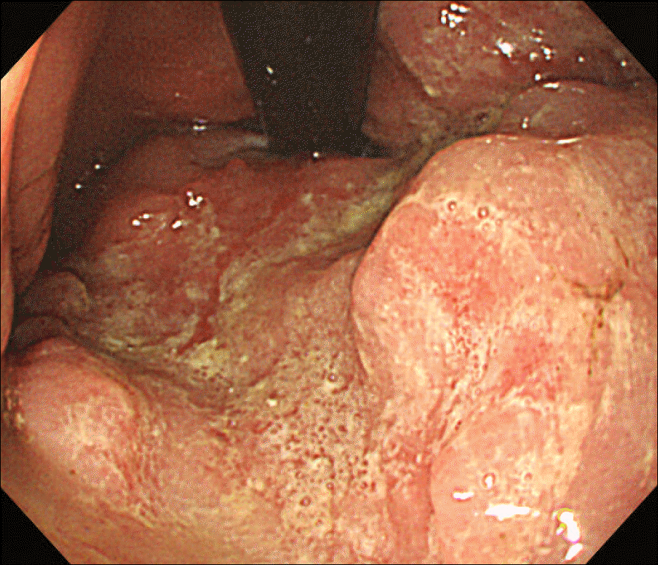
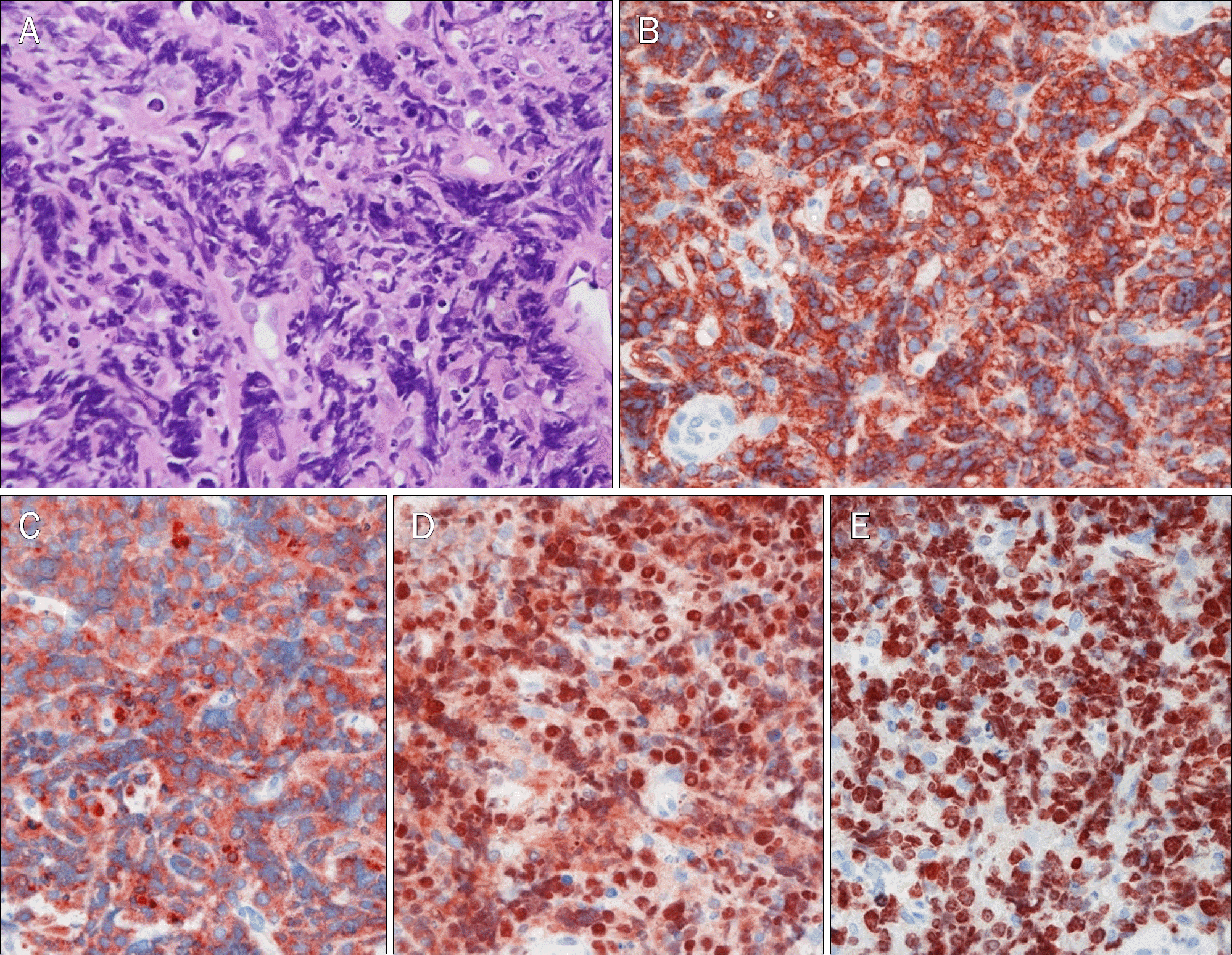
Fig. 4.
Gastric tissue histology. (A) This slide shows lymphoid and monomorphic round cells is diffusely infiltrated within mucosa (H&E, ×400). The immunohistochemistry slides show the destructive infiltration of B-cell lineage. The tumor cells are positive for (B) CD20 (×400), (C) CD10 (×400), and (D) Bcl-6 protein (×400). (E) Total tumor cells are Ki-67 positive (×400).


 PDF
PDF ePub
ePub Citation
 Print
Print


 XML Download