Abstract
Background/Aims
Peptic ulcer bleeding (PUB) is the most common cause of upper gastrointestinal bleeding in Korea but there has been no research done using big data. This study evaluates the optimal operational definition (OD) for big data research by analyzing clinical characteristics of PUB.
Methods
We reviewed the clinical characteristics of 92 patients with PUB confirmed on endoscopy in Wonkwang University Sanbon Hospital (January 2013 to December 2014). We calculated sensitivity and positive predictive value (PPV) to detect confirmed PUB patients using ODs developed by combining clinical features of patients with PUB.
Results
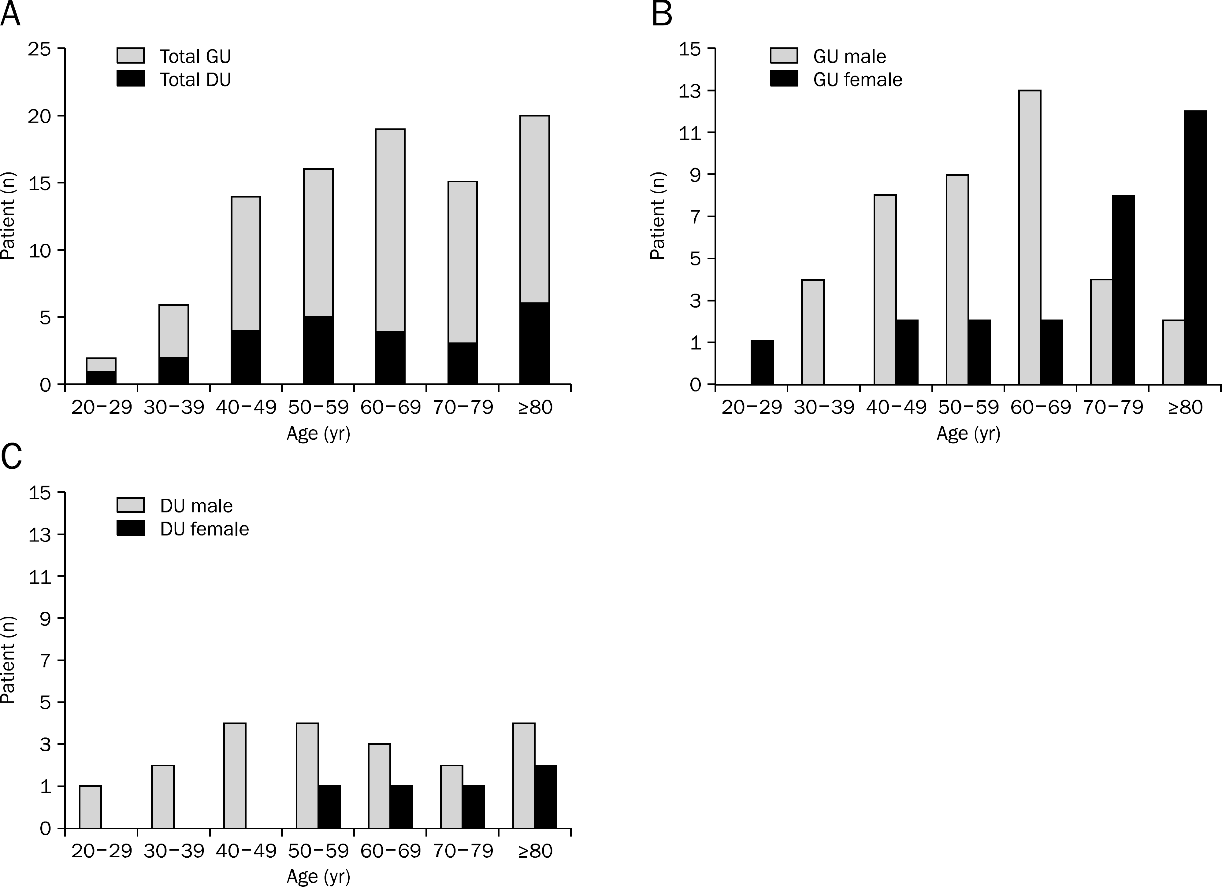
The mean patient age was 63 years. Men had higher prevalence of PUB than women. Bleeding gastric ulcer was proportionately common in the age range of 40s to 60s in men, while a significantly higher rate of bleeding occurred in women older than 70s. The rate of drug-induced ulcer was 28.2%, whereas the prevalence of Helicobacter pylori was 47.8%. Among the hospitalized patients with diagnostic code of PUB, we ruled out patients with endoscopic removal of gastric adenoma or peritonitis, and selected patients who had been administered intravenous proton pump inhibitor. The sensitivity in this setting was 82.6%, and PPV was 88.4%.
Go to : 
References
3. Wu CY, Wu CH, Wu MS, et al. A nationwide population-based cohort study shows reduced hospitalization for peptic ulcer disease associated with H pylori eradication and proton pump inhibitor use. Clin Gastroenterol Hepatol. 2009; 7:427–431.
4. Ahsberg K, Ye W, Lu Y, Zheng Z, Staël von Holstein C. Hospitalisation of and mortality from bleeding peptic ulcer in Sweden: a nationwide time-trend analysis. Aliment Pharmacol Ther. 2011; 33:578–584.
5. Theocharis GJ, Thomopoulos KC, Sakellaropoulos G, Katsakoulis E, Nikolopoulou V. Changing trends in the epidemiology and clinical outcome of acute upper gastrointestinal bleeding in a defined geographical area in Greece. J Clin Gastroenterol. 2008; 42:128–133.

6. Kwon JH, Choi MG, Lee SW, et al. Trends of gastrointestinal diseases at a single institution in Korea over the past two decades. Gut Liver. 2009; 3:252–258.

7. Nagasue T, Nakamura S, Kochi S, et al. Time trends of the impact of Helicobacter pylori infection and nonsteroidal anti-inflammatory drugs on peptic ulcer bleeding in Japanese patients. Digestion. 2015; 91:37–41.
8. Choi JW, Kim HY, Kim KH, et al. Has any improvement been made in the clinical outcome of patients with bleeding peptic ulcer in the part 10 years? Korean J Gastrointest Endosc. 2005; 30:235–242.
9. Kim JJ, Kim N, Lee BH, et al. Risk factors for development and recurrence of peptic ulcer disease. Korean J Gastroenterol. 2010; 56:220–228.

10. Na YJ, Shim KN, Kang MJ, et al. Comparison of clinical characteristics and outcomes between geriatric and non-geriatric patients in peptic ulcer bleeding. Korean J Gastroenterol. 2009; 53:297–304.

11. Bae SO, Kang GW. A comparative study of the disease codes between Korean national health insurance vlaims and Korean national hospital discharge in-depth injury survey. Health Policy Manag. 2014; 24:322–329.
12. Bae S, Kim N, Kang JM, et al. Incidence and 30-day mortality of peptic ulcer bleeding in Korea. Eur J Gastroenterol Hepatol. 2012; 24:675–682.

13. Kim JJ, Kim N, Park HK, et al. Clinical characteristics of patients diagnosed as peptic ulcer disease in the third referral center in 2007. Korean J Gastroenterol. 2012; 59:338–346.

14. Huang JQ, Sridhar S, Hunt RH. Role of Helicobacter pylori infection and non-steroidal anti-inflammatory drugs in peptic-ulcer disease: a metaanalysis. Lancet. 2002; 359:14–22.
15. Lim SH, Kwon JW, Kim N, et al. Prevalence and risk factors of Helicobacter pylori infection in Korea: nationwide multicenter study over 13 years. BMC Gastroenterol. 2013; 13:104.

16. Güell M, Artigau E, Esteve V, Sánchez-Delgado J, Junquera F, Calvet X. Usefulness of a delayed test for the diagnosis of Helicobacter pylori infection in bleeding peptic ulcer. Aliment Pharmacol Ther. 2006; 23:53–59.
17. Ko MJ, Lim TH. Use of big data for evidence-based healthcare. J Korean Med Assoc. 2014; 57:413–418.

19. Gilbert DA, Saunders DR. Iced saline lavage does not slow bleeding from experimental canine gastric ulcers. Dig Dis Sci. 1981; 26:1065–1068.

Go to : 
 | Fig. 1.Distribution by age and sex. (A) Patients with peptic ulcer bleeding were evenly distributed over the forties age decade. (B) In gender analysis of gastric ulcer (GU) bleeding, the proportions of patients with bleeding gastric ulcer was high in the age range of 40s to 60s in men, while higher rate of bleeding occurred at age older than 70s in female (p<0.001, Fisher's exact test). (C) There was no significant gender difference in duodenal ulcer (DU) bleeding. |
Table 1.
Diagnostic Codes for Peptic Ulcer Disease Used in This Study
| Code a | Disease |
|---|---|
| Diagnostic codes for peptic ulcer disease including hemorrhage | |
| K25.0 | Acute gastric ulcer with hemorrhage |
| K25.2 | Acute gastric ulcer with both hemorrhage and perforation |
| K25.4 | Chronic or unspecified gastric ulcer with hemorrhage |
| K25.6 | Chronic or unspecified gastric ulcer with both hemorrhage and perforation |
| K26.0 | Acute duodenal ulcer with hemorrhage |
| K26.2 | Acute duodenal ulcer with both hemorrhage and perforation |
| K26.4 | Chronic or unspecified duodenal ulcer with hemorrhage |
| K26.6 | Chronic or unspecified duodenal ulcer with both hemorrhage and perforation |
| K27.0 | Acute peptic ulcer, site unspecified with hemorrhage |
| K27.2 | Acute peptic ulcer, site unspecified with both hemorrhage and perforation |
| K27.4 | Chronic or unspecified peptic ulcer, site unspecified with hemorrhage |
| K27.6 | Chronic or unspecified peptic ulcer, site unspecified with both hemorrhage and perforation |
| Diagnostic codes for peptic ulcer disease without complication | |
| K25 | Gastric ulcer |
| K25.3 | Acute gastric ulcer without hemorrhage or perforation |
| K25.7 | Chronic gastric ulcer without hemorrhage or perforation |
| K25.9 | Unspecified as acute or chronic gastric ulcer without hemorrhage or perforation |
| K26 | Duodenal ulcer |
| K26.3 | Acute duodenal ulcer without hemorrhage or perforation |
| K26.7 | Chronic duodenal ulcer without hemorrhage or perforation |
| K26.9 | Unspecified as acute or chronic duodenal ulcer without hemorrhage or perforation |
| K27 | Peptic ulcer, site unspecified |
| K27.3 | Acute peptic ulcer, site unspecified without hemorrhage or perforation |
| K27.7 | Chronic peptic ulcer, site unspecified without hemorrhage or perforation |
| K27.9 | Unspecified as acute or chronic peptic ulcer, site unspecified without hemorrhage or perforation |
Table 2.
Demographic Data of Patients with Bleeding Peptic Ulcer Disease
| Total | Male | Female | p-value c | |
|---|---|---|---|---|
| Number of patients | 92 (100) | 60 (65.2) | 32 (34.8) | |
| Age (yr) | 63±16.7 | 58±14.7 | 73±15.7 | <0.001 d |
| Location of ulcer | NS e | |||
| GU | 64 (69.6) | 38 a (63.3) | 26 (81.3) | |
| DU | 19 (20.7) | 16 (26.7) | 3 (9.4) | |
| GU+DU | 9 (9.8) | 6 (10.0) | 3 (9.4) | |
| Bleeding site | NS f | |||
| GU | 67 (72.8) | 40 (66.7) | 27 (84.4) | |
| DU | 25 (27.2) | 20 (33.3) | 5 (15.6) | |
| Stage of ulcer | NS f | |||
| A1 | 32 (34.8) | 24 (40.0) | 8 (25.0) | |
| A2 | 46 (50.0) | 30 (50.0) | 16 (50.0) | |
| H1 | 12 (13.0) | 5 (8.3) | 7 (21.9) | |
| H2 | 2 (2.2) | 1 (1.7) | 1 (3.1) | |
| Forrest classification | <0.01 f | |||
| IA | 5 (5.4) | 4 (6.7) | 1 (3.1) | |
| IB | 5 (5.4) | 5 (8.3) | 0 (0.0) | |
| IIA | 16 (17.4) | 14 (23.3) | 2 (6.3) | |
| IIB | 6 (6.5) | 4 (6.7) | 2 (6.3) | |
| IIC | 40 (43.5) | 26 (43.3) | 14 (43.8) | |
| III | 20 (21.7) | 7 (11.7) | 13 (40.6) | |
| Drug history b | 26 (28.2) | 17 (28.3) | 9 (28.1) | NS f |
| NSAIDS | 15 (16.3) | 10 (16.7) | 5 (15.6) | |
| Aspirin | 5 (5.4) | 2 (3.3) | 3 (9.4) | |
| Steroid | 5 (5.4) | 2 (3.3) | 3 (9.4) | |
| Anti-platelet agent | 9 (9.8) | 7 (11.7) | 2 (6.3) |
Table 3.
Hemostasis Method
Table 4.
Infection Rate and Diagnostic Method for Helicobacter pylori
Table 5.
Operational Definition Combinations
| Combinations | Total patients (n) | Confirmed (n) | Not confirmed (n) | Sensitivity a (%) | PPV (%) |
|---|---|---|---|---|---|
| Combination A (operational definition used in previous study12) | |||||
| 1. All PUD code AND [Hemostasis code or EGD with L-tube insertion code] | 61 | 37 | 24 | 40.2 | 60.7 |
| 2. All PUD code AND [Hemostasis code or EGD with L-tube insertion code] AND IV PPI | 59 | 37 | 22 | 40.2 | 62.7 |
| Combination B (coding during admission period only for new onset and recurrent cases) | |||||
| 1. PUD code including hemorrhage | 99 | 65 | 34 | 70.7 | 65.7 |
| 2. PUD code including hemorrhage AND Exclusion of EMR/ESD/peritonitis code | 88 | 65 | 23 | 70.7 | 73.9 |
| 3. PUD code including hemorrhage AND Exclusion of EMR/ESD/peritonitis code AND IV PPI | 75 | 65 | 10 | 70.7 | 86.7 |
| Combination C (coding during admission period and within 1 month after discharge b for new onset and recurrent cases) | |||||
| 1. PUD code including hemorrhage | 107 | 73 | 34 | 79.3 | 68.2 |
| 2. PUD code including hemorrhage AND Exclusion of EMR/ESD/peritonitis code | 96 | 73 | 23 | 79.3 | 76.0 |
| 3. PUD code including hemorrhage AND Exclusion of EMR/ESD/peritonitis code AND IV PPI | 83 | 73 | 10 | 79.3 | 88.0 |
| Combination D (coding before, during admission period, and within 1 month after discharge for new onset and recurrent cases) | |||||
| 1. PUD code including hemorrhage | 117 | 76 | 41 | 82.6 | 65.0 |
| 2. PUD code including hemorrhage AND Exclusion of EMR/ESD/peritonitis code | 105 | 76 | 29 | 82.6 | 72.4 |
| 3. PUD code including hemorrhage AND Exclusion of EMR/ESD/peritonitis code AND IV PPI | 86 | 76 | 10 | 82.6 | 88.4 |
| Combination E (add combinations using codes of PUD without complication to combination D for new onset and recurrent cases) | |||||
| 1. Combination D-3+[PUD code without complication AND Hemostasis code AND Exclusion of EMR/ESD code] | 89 | 78 | 11 | 84.8 | 87.6 |
| 2. Combination D-3+[PUD code without complication AND Hemostasis code with Exclusion of EMR/ESD code]+[PUD code without complication AND EGD with L-tube insertion/IV PPI] | 94 | 79 | 15 | 85.9 | 84.0 |
| Combination F (coding during admission period and within 1month after discharge for new onset cases only) | |||||
| 1. PUD code including hemorrhage AND Exclusion of EMR/ESD/peritonitis code AND IV PPI | 83 | 73 | 10 | 82.0 | 88.0 |
| 2. Combination F-1+[PUD code without complication AND Hemostasis code AND Exclusion of EMR/ESD code] | 86 | 75 | 11 | 84.3 | 87.2 |
| 3. Combination F-1+[PUD code without complication AND Hemostasis code with Exclusion of EMR/ESD code]+[PUD code without complication AND EGD with L-tube insertion/IV PPI] | 91 | 76 | 15 | 85.4 | 83.5 |
PUD, peptic ulcer disease; EGD, esophagogastroduodenoscopy; IV, intravenous; PPI, proton pump inhibitor; PPV, positive predictive value
a Validation is on the basis of a cohort of 92 true patients with bleeding PUD who were confirmed by upper endoscopy in Wonkwang University Sanbon Hospital during 2013–2014. For sensitivity calculation, the number of true patients was 92 for new onset and recurrent bleeding PUD cases and 89 for new onset bleeding PUD cases only


 PDF
PDF ePub
ePub Citation
 Print
Print


 XML Download