Abstract
Many neoplasms, including lung cancer, breast cancer, melanoma, and gastrointestinal tract malignancy, possess potential for skin metastasis. Skin metastases can represent the first presentation of such malignancies and may be observed incidentally during routine exam. Skin metastases from gastric adenocarcinoma are uncommon, with a prevalence rate of 0.04–0.8%. Cutaneous metastases from gastric cancer are generally observed as the initial symptom of advanced gastric cancer. Early detection and treatment can increase patient survival. A 42-year-old woman visited our department with nodule about 1 cm in size on the right frontal scalp noticed incidentally after laparoscopy-assisted distal gastrectomy and adjuvant systemic chemotherapy for early gastric cancer about 16 months prior. The patient was diagnosed with skin metastasis from gastric adenocarcinoma. Complete excision of the skin lesion and additional chemotherapy were performed. Herein, we report a case of nodular tumor-like scalp metastasis from early gastric cancer with a brief review of the literature.
References
1. Hu SC, Chen GS, Wu CS, Chai CY, Chen WT, Lan CC. Rates of cutaneous metastases from different internal malignancies: experience from a Taiwanese medical center. J Am Acad Dermatol. 2009; 60:379–387.

2. Lookingbill DP, Spangler N, Helm KF. Cutaneous metastases in patients with metastatic carcinoma: a retrospective study of 4020 patients. J Am Acad Dermatol. 1993; 29:228–236.

3. Saeed S, Keehn CA, Morgan MB. Cutaneous metastasis: a clinical, pathological, and immunohistochemical appraisal. J Cutan Pathol. 2004; 31:419–430.

4. Kim YC, Cho KH, Lee YS, Ham EK. Cutaneous metastasis from internal malignancy. Korean J Dermatol. 1987; 25:213–221.
5. Lee SY, Hahn CY, Lee JF, et al. MAOA interacts with the ALDH2 gene in anxiety-depression alcohol dependence. Alcohol Clin Exp Res. 2010; 34:1212–1218.

6. Jang YH, Lim do H, Kim YH, et al. Early gastric cancer with celluli-tis-like skin metastasis. Korean J Gastroenterol. 2014; 63:39–41.

7. Frey L, Vetter-Kauczok C, Gesierich A, Bröcker EB, Ugurel S. Cutaneous metastases as the first clinical sign of metastatic gastric carcinoma. J Dtsch Dermatol Ges. 2009; 7:893–895.

8. Lifshitz OH, Berlin JM, Taylor JS, Bergfeld WF. Metastatic gastric adenocarcinoma presenting as an enlarging plaque on the scalp. Cutis. 2005; 76:194–196.
9. Kim HJ, Min HG, Lee ES. Alopecia neoplastica in a patient with gastric carcinoma. Br J Dermatol. 1999; 141:1122–1124.

10. Fernández-Antón Martínez MC, Parra-Blanco V, Avilés Izquierdo JA, Suárez Fernández RM. Cutaneous metastases of internal tumors. Actas Dermosifiliogr. 2013; 104:841–853.
11. Michiwa Y, Earashi M, Kobayashi H, Matsuki N. Cutaneous metastases from gastric adenocarcinoma treated with combination chemotherapy producing complete response with long survival. J Exp Clin Cancer Res. 2001; 20:297–299.
12. Cesaretti M, Malerba M, Basso V, et al. Cutaneous metastasis from primary gastric cancer: a case report and review of the literature. Cutis. 2014; 93:E9–E13.
Fig. 1.
Upper endoscopic finding taken in its initial presentation. Endoscopy shows a reddish lesion, about 2 cm, with irregular surface and central shallow depression on the gastric antrum.

Fig. 2.
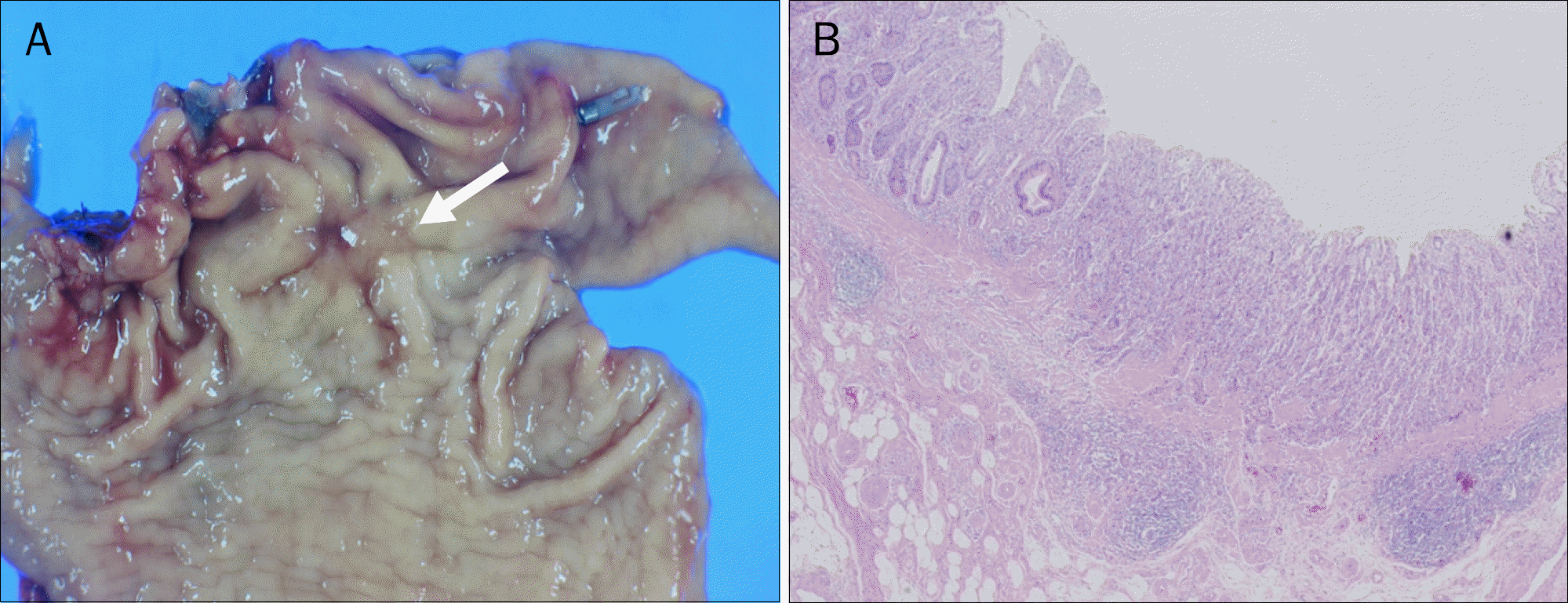
Resected stomach pathologic findings. (A) The arrow indicates gross finding at early gastric cancer site. (B) Poorly differentiated tubular adenocarcinoma have invaded the submucosal layer, above 500 μm (H&E, ×100).


 PDF
PDF ePub
ePub Citation
 Print
Print


 XML Download