

 PDF
PDF ePub
ePub Citation
 Print
Print


Abstract
Oral metastatic tumor, which is uncommon and represents less than 1% of malignant oral neoplasms, usually arises from a primary mucosal or cutaneous cancer located in the head and neck regions. Metastasis of hepatocellular carcinoma (HCC) to the oral cavity, especially to gingiva, is extremely rare. A 50-year-old man, who was a chronic alcoholic and hepatitis B virus carrier, presented with abdominal distension and weight loss for the past 3 months. Three-phased contrast-enhanced abdominal CT revealed numerous conglomerated masses in the liver, suggesting huge HCCs arising in the background of liver cirrhosis with a large amount of ascites. He complained of recurrent profuse bleeding from the left upper gingival mass. A facial CT revealed an oral cavity mass destructing the left maxillary alveolar process and hard palate, which was diagnosed as metastatic HCC by an incisional biopsy. Herein, we report a case of metastatic HCC to the gingiva.
Go to : 
References
1. Kao JH, Chen DS. Changing disease burden of hepatocellular carcinoma in the Far East and Southeast Asia. Liver Int. 2005; 25:696–703.

2. Huang SF, Wu RC, Chang JT, et al. Intractable bleeding from solitary mandibular metastasis of hepatocellular carcinoma. World J Gastroenterol. 2007; 13:4526–4528.
3. Sawabe M, Nakamura T, Kanno J, Kasuga T. Analysis of morphological factors of hepatocellular carcinoma in 98 autopsy cases with respect to pulmonary metastasis. Acta Pathol Jpn. 1987; 37:1389–1404.
4. Katyal S, Oliver JH 3rd, Peterson MS, Ferris JV, Carr BS, Baron RL. Extrahepatic metastases of hepatocellular carcinoma. Radiology. 2000; 216:698–703.
5. Natsuizaka M, Omura T, Akaike T, et al. Clinical features of hepatocellular carcinoma with extrahepatic metastases. J Gastroenterol Hepatol. 2005; 20:1781–1787.
6. Watanabe J, Nakashima O, Kojiro M. Clinicopathologic study on lymph node metastasis of hepatocellular carcinoma: a retrospective study of 660 consecutive autopsy cases. Jpn J Clin Oncol. 1994; 24:37–41.
7. Ramón Ramirez J, Seoane J, Montero J, Esparza Gómez GC, Cerero R. Isolated gingival metastasis from hepatocellular carcinoma mimicking a pyogenic granuloma. J Clin Periodontol. 2003; 30:926–929.
8. Lee YT, Geer DA. Primary liver cancer: pattern of metastasis. J Surg Oncol. 1987; 36:26–31.
9. Yoshimura Y, Matsuda S, Naitoh S. Hepatocellular carcinoma metastatic to the mandibular ramus and condyle: report of a case and review of the literature. J Oral Maxillofac Surg. 1997; 55:297–306.
10. Pires FR, Sagarra R, Corrêa ME, Pereira CM, Vargas PA, Lopes MA. Oral metastasis of a hepatocellular carcinoma. Oral Surg Oral Med Oral Pathol Oral Radiol Endod. 2004; 97:359–368.
11. Will TA, Agarwal N, Petruzzelli GJ. Oral cavity metastasis of renal cell carcinoma: a case report. J Med Case Rep. 2008; 2:313.
12. Makos CP, Psomaderis K. A literature review in renal carcinoma metastasis to the oral mucosa and a new report of an epulis-like metastasis. J Oral Maxillofac Surg. 2009; 67:653–660.
13. Shin SJ, Roh JL, Choi SH, et al. Metastatic carcinomas to the oral cavity and oropharynx. Korean J Pathol. 2012; 46:266–271.
14. Batson OV. The function of the vertebral veins and their role in the spread of metastases. Ann Surg. 1940; 112:138–149.
15. Morishita M, Fukuda J. Hepatocellular carcinoma metastatic to the maxillary incisal gingiva. J Oral Maxillofac Surg. 1984; 42:812–815.
16. Gong LI, Zhang WD, Mu XR, et al. Hepatocellular carcinoma metastasis to the gingival soft tissues: a case report and review of the literature. Oncol Lett. 2015; 10:1565–1568.
17. Hwang SW, Lee JE, Lee JM, et al. Hepatocellular carcinoma with cervical spine and pelvic bone metastases presenting as unknown primary neoplasm. Korean J Gastroenterol. 2015; 66:50–54.
18. Inaba H, Kanazawa N, Wada I, et al. A case of hepatocellular carcinoma with bleeding gingival metastasis treated by transcatheter arterial embolization. Nihon Shokakibyo Gakkai Zasshi. 2011; 108:95–102.
Go to :
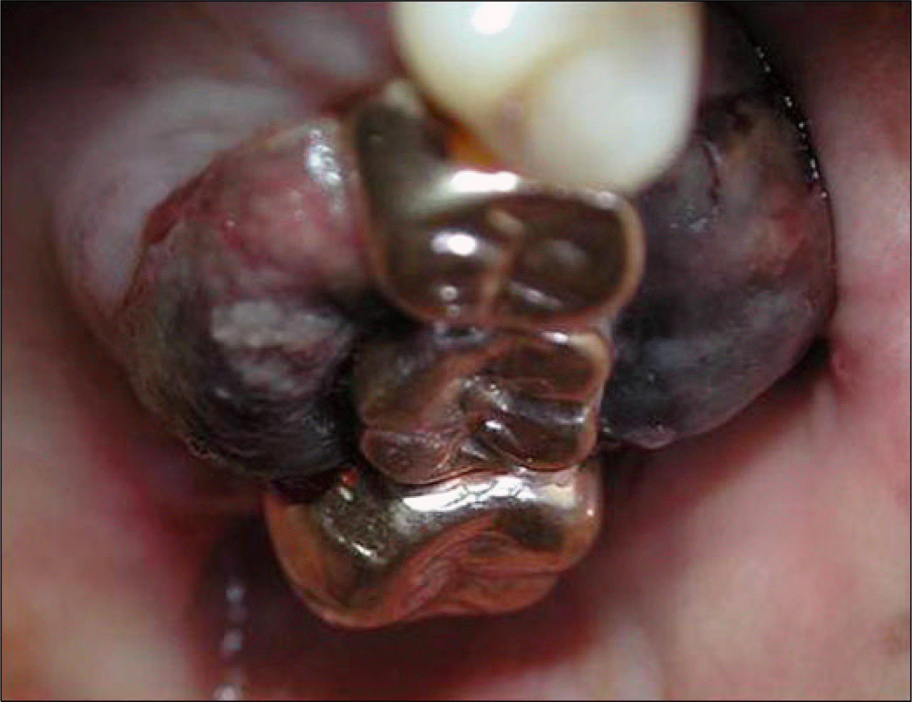 | Fig. 1.A protruding mass suggesting metastatic hepatocellular carcinoma is noted at the left gingiva. |
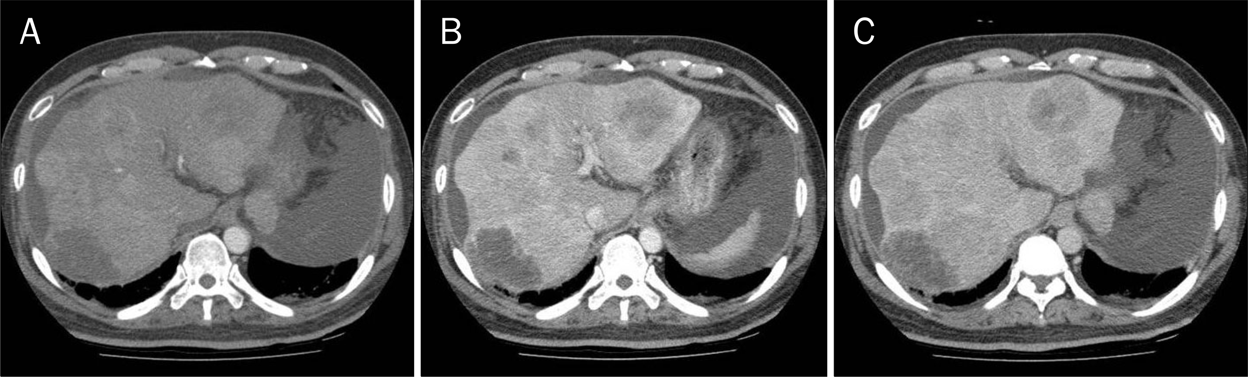 | Fig. 2.(A) Arterial phase, (B) portal phase, (C) delayed phase. Numerous hepatocellular carcinomas are seen in the entire liver on abdominal CT. These masses show heterogeneous enhancement in arterial phase and typical wash-out pattern in portal and delayed phase. |
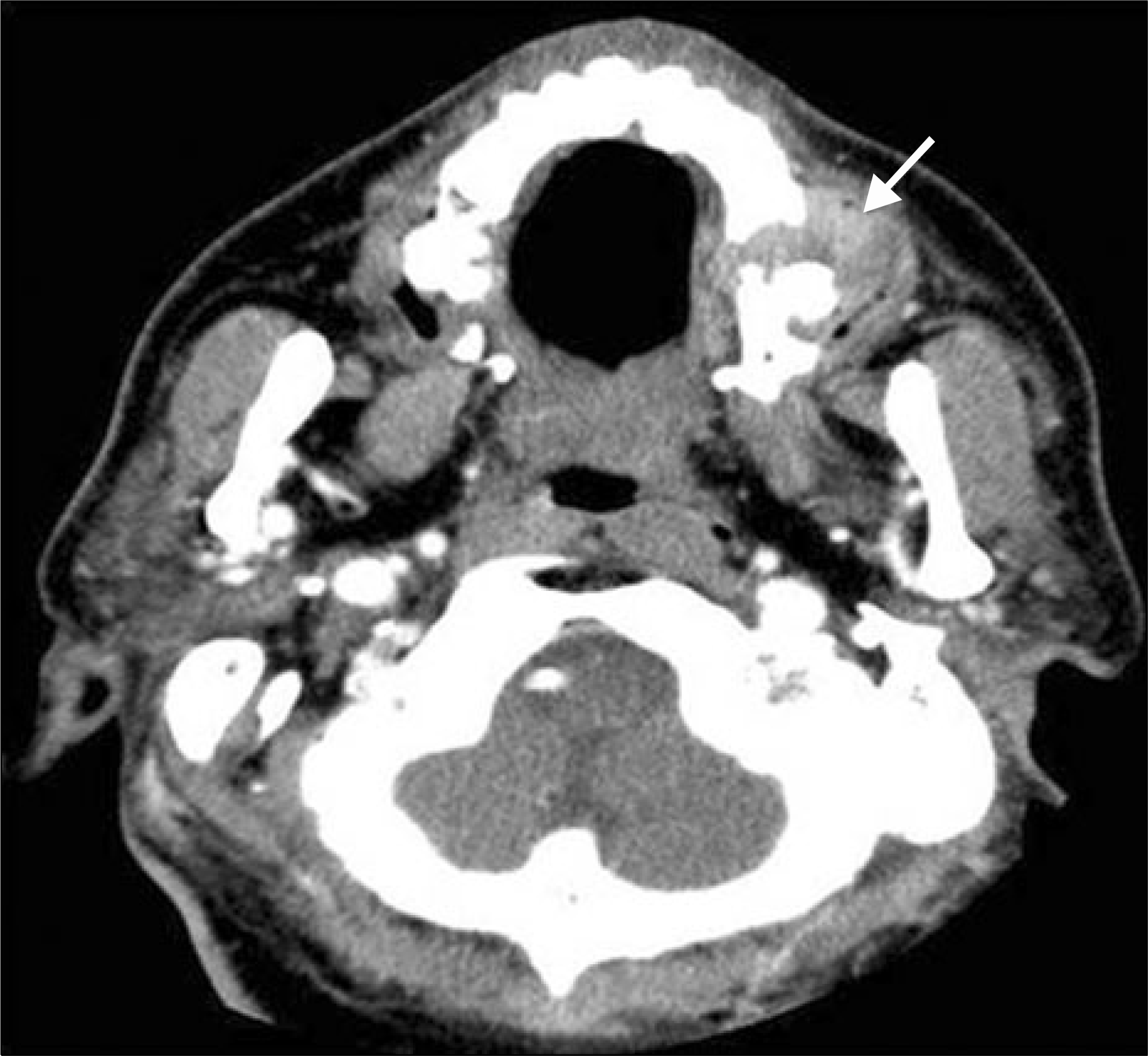 | Fig. 3.Facial CT presents malignant tumor involving the alveolar process of the left maxilla and lateral aspect of the left side hard palate (arrow). |
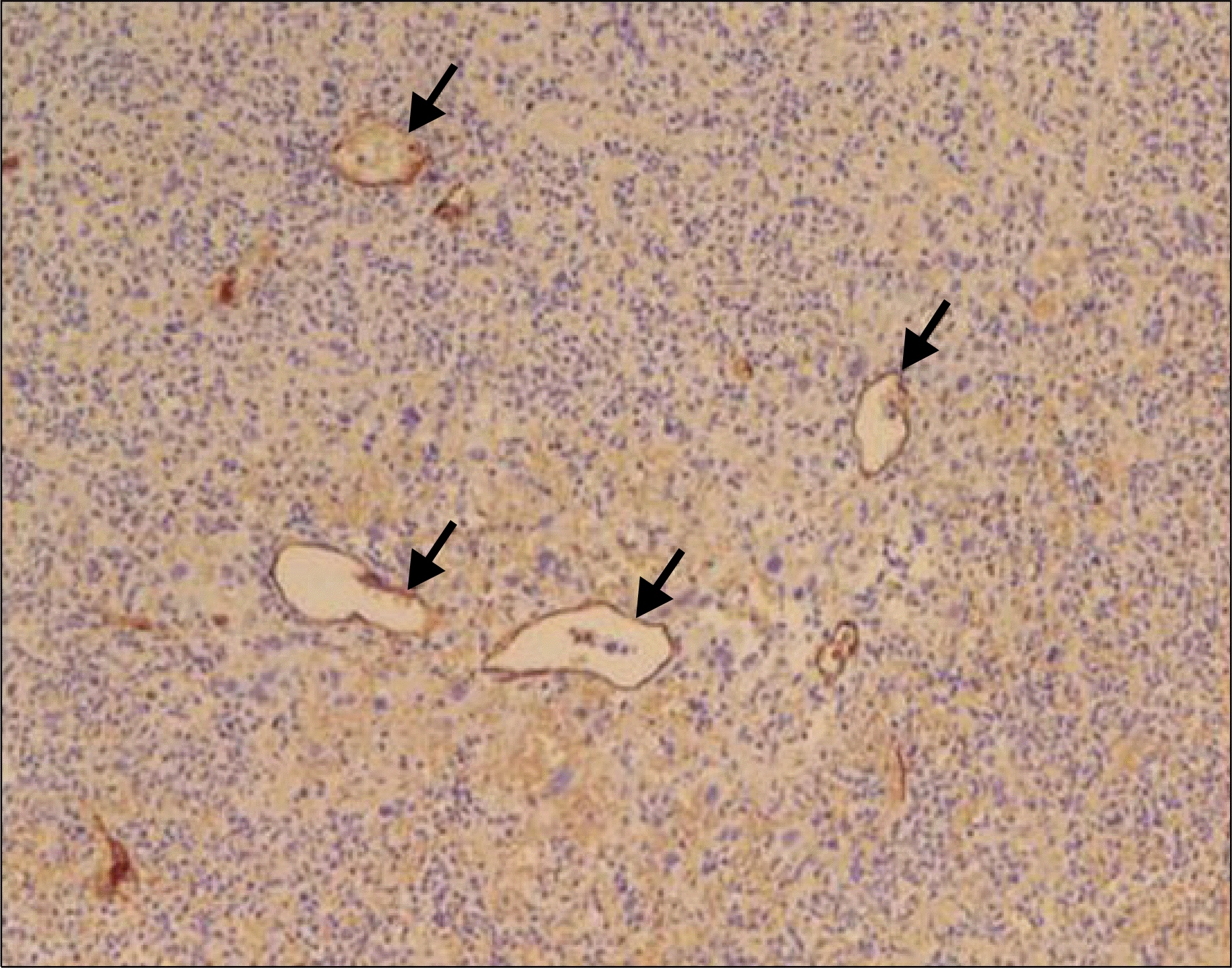
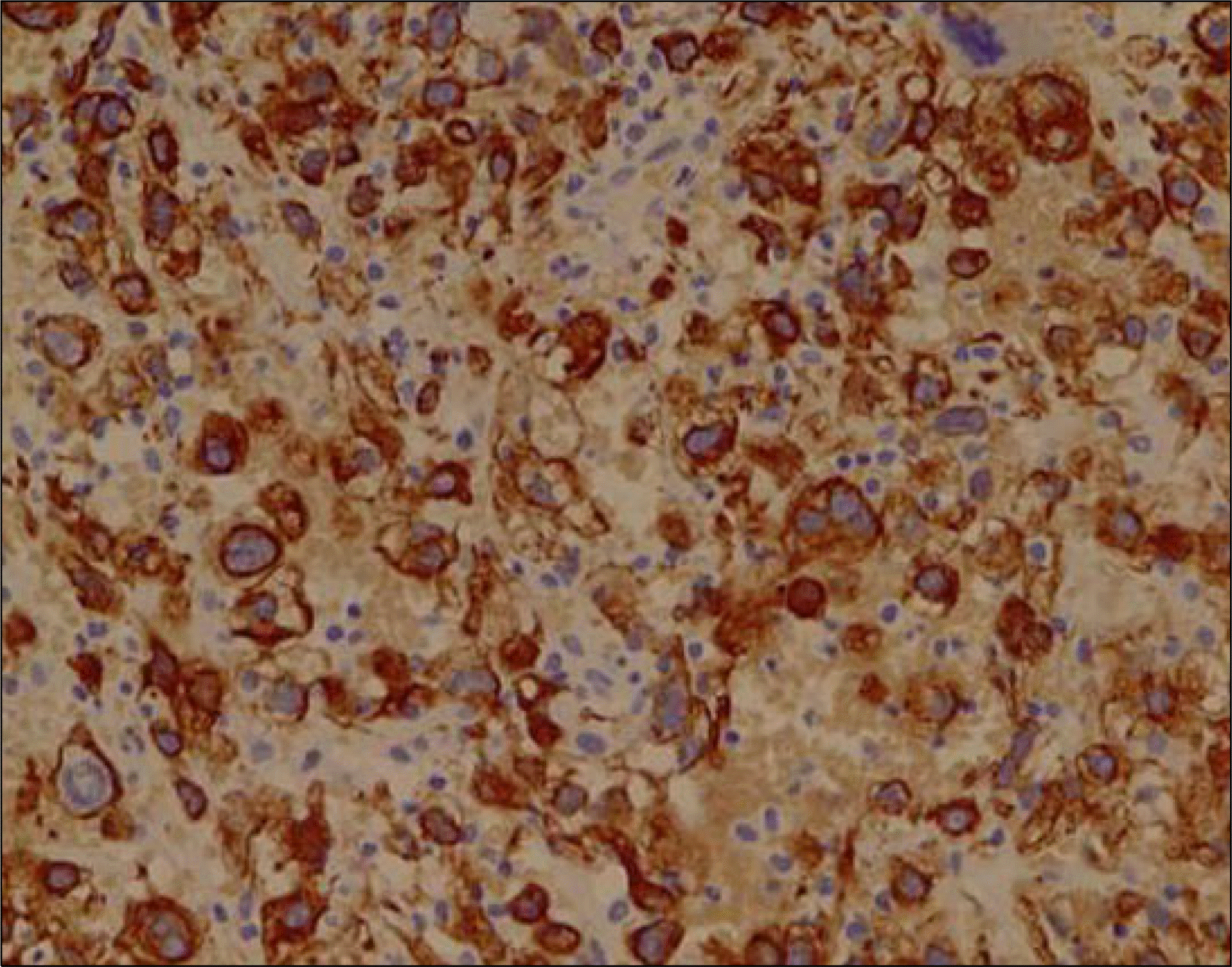
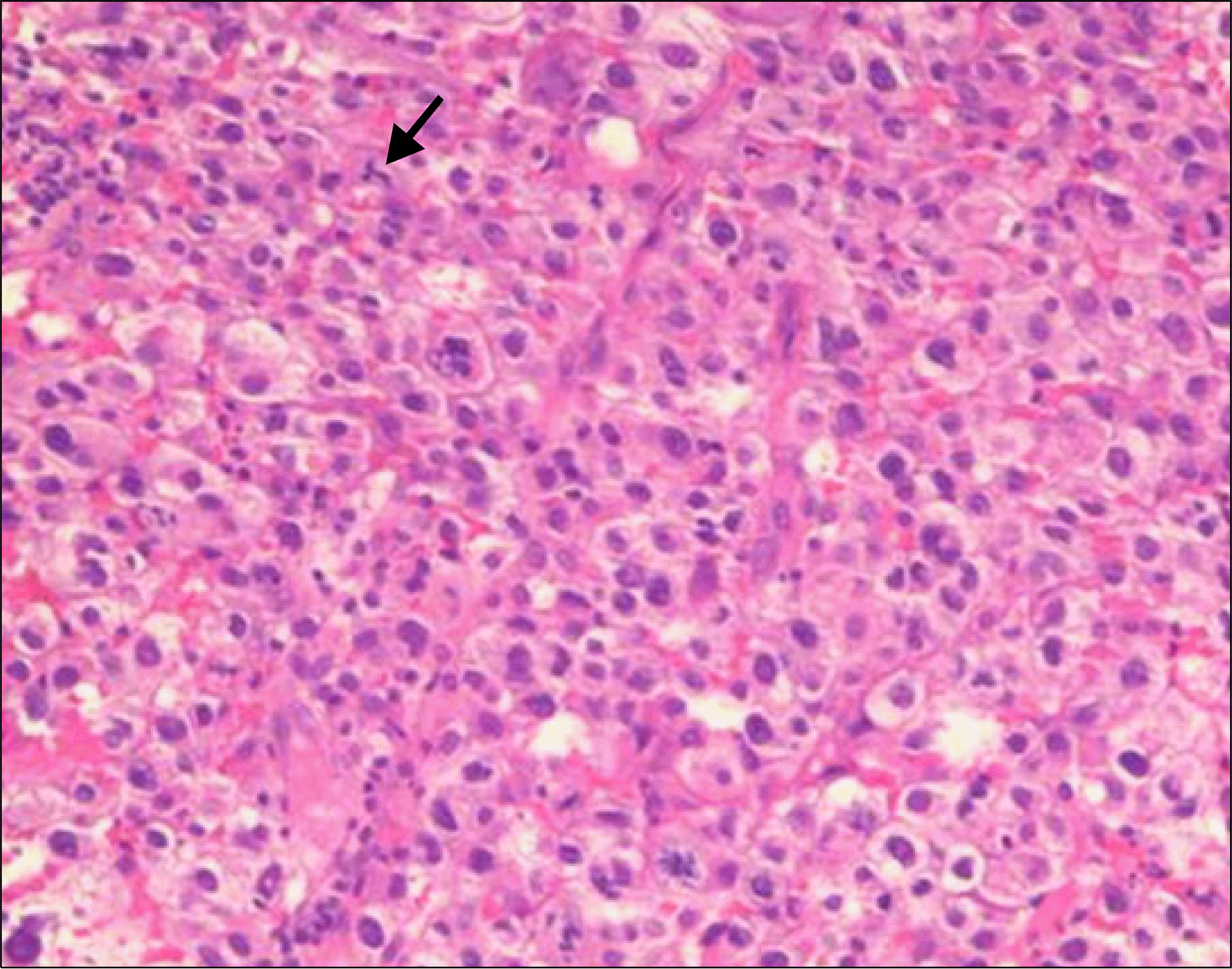 | Fig. 4.Photomicrograph of oral mass exhibiting severe nuclear anaplasia and pleomorphism, frequent mitotic figures, including atypical tripolar mitotic spindle (arrow) and sheet to trabecular arrangement. H&E stain, ×200. |
 XML Download