

 PDF
PDF ePub
ePub Citation
 Print
Print


Abstract
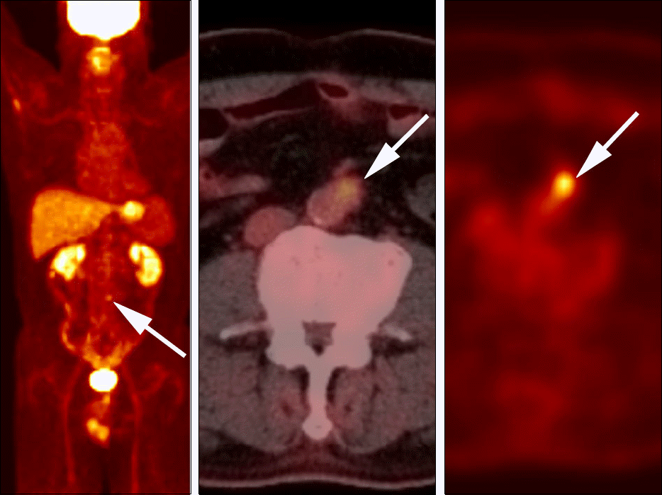
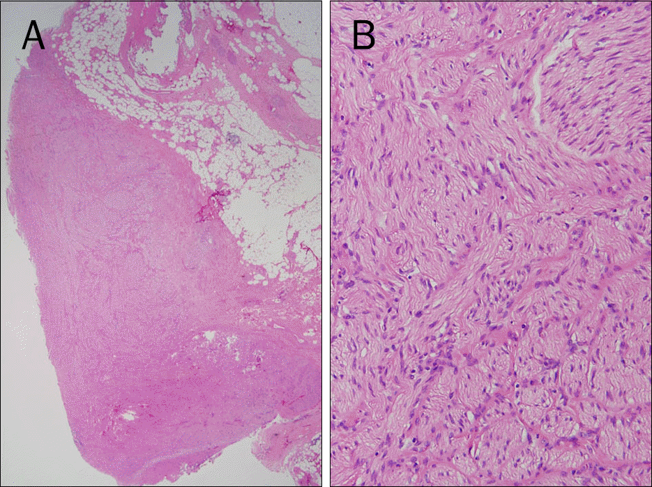
Traumatic neuroma results from regeneration attempts of the proximal end of an injured or severed nerve, resulting in a non-neoplastic nodular lesion. The lower extremity after amputation is the most common site, followed by the head and neck. Traumatic neuromas occurring in the abdomen, however, are rare. In the abdominal region, traumatic neuromas occur in the cystic duct stump and the common bile ducts as well as around the celiac trunk. This study reports a case of a 59-year-old man who presented with a traumatic neuroma arising at the stump of the inferior mesenteric artery after rectal cancer surgery. Traumatic neuromas at the stump of the inferior mesenteric artery have not been previously reported. The lesion exhibited atypical imaging features, including a well-enhanced nodule, a significant interval growth in size and a mild increase in 18F-fluo-rodeoxyglucose uptake, resembling lymph node metastasis. This case report will help physicians understand the sites of occurrence and imaging features of traumatic neuromas in the abdomen.
Go to : 
References
1. Huang LF, Weissman JL, Fan C. Traumatic neuroma after neck dissection: CT characteristics in four cases. AJNR Am J Neuroradiol. 2000; 21:1676–1680.
2. Yabuuchi H, Kuroiwa T, Fukuya T, Tomita K, Hachitanda Y. Traumatic neuroma and recurrent lymphadenopathy after neck dissection: comparison of radiologic features. Radiology. 2004; 233:523–529.

3. Boutin RD, Pathria MN, Resnick D. Disorders in the stumps of amputee patients: MR imaging. AJR Am J Roentgenol. 1998; 171:497–501.
4. Brogan DM, Kakar S. Management of neuromas of the upper extremity. Hand Clin. 2013; 29:409–420.
5. Swanson HH. Traumatic neuromas. A review of the literature. Oral Surg Oral Med Oral Pathol. 1961; 14:317–326.
6. Murphey MD, Smith WS, Smith SE, Kransdorf MJ, Temple HT. From the archives of the AFIP. Imaging of musculoskeletal neurogenic tumors: radiologic-pathologic correlation. Radiographics. 1999; 19:1253–1280.
7. Nagata Y, Tomioka T, Chiba K, Kanematsu T. Traumatic neuroma of the common hepatic duct after laparoscopic cholecystectomy. Am J Gastroenterol. 1995; 90:1887–1888.
8. Koh DW, Lee WJ, Kim JH, et al. Amputation neuroma mimicking common bile duct cancer: a case report. Korean J Gastroenterol. 2008; 52:32–36.
9. Shimura K, Tamada K, Asada M, et al. Intraductal ultrasonography of traumatic neuroma of the bile duct. Abdom Imaging. 2001; 26:632–634.
10. van Gulik TM, Brummelkamp WH, Lygidakis NJ. Traumatic neuroma giving rise to biliary obstruction after reconstructive surgery for iatrogenic lesions of the biliary tract–a report of three cases. Hepatogastroenterology. 1989; 36:255–257.
11. Mentha G, Rubbia-Brandt L, Orci L, et al. Traumatic neuroma with biliary duct obstruction after orthotopic liver transplantation. Transplantation. 1999; 67:177–179.
12. Katsinelos P, Dimiropoulos S, Galanis I, et al. Biliary stricture due to neuroma after an innocent blunt abdominal trauma. Surg Endosc. 2002; 16:1494.
13. Kwon JH, Ryu SW, Kang YN. Traumatic neuroma around the celiac trunk after gastrectomy mimicking a nodal metastasis: a case report. Korean J Radiol. 2007; 8:242–245.
14. Curran T, Poylin V, Kane R, Harris A, Goldsmith JD, Nagle D. Case report of a traumatic rectal neuroma. Gastroenterol Rep (Oxf). 2015. DOI: doi:10.1093/gastro/gov023.
15. Ahlawat S, Belzberg AJ, Montgomery EA, Fayad LM. MRI features of peripheral traumatic neuromas. Eur Radiol. 2016; 26:1204–1212.
16. Chang JM, Lee HJ, Goo JM, et al. False positive and false negative FDG-PET scans in various thoracic diseases. Korean J Radiol. 2006; 7:57–69.
Go to :
 | Fig. 1.An ill-defined left para-aortic lesion on CT images obtained two weeks after surgery. (A) Non-contrast CT reveals an ill-defined infiltrative para-aortic soft tissue attenuation lesion (arrow, 30.53±27.25 HU) at the inferior mesenteric artery stump area. (B) Portal-venous phase CT indicates that the lesion (arrow) is persistently enhancing (53.64±28.65 HU). This lesion was interpreted as a postoperative granulation tissue because the occurrence of a lesion at a stump is considered a typical finding. |
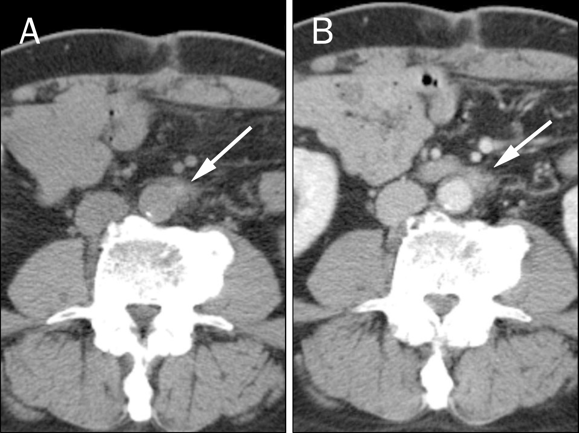
 | Fig. 2.A left para-aortic lesion changing into a discrete nodule on follow-up CT scans. (A) Portal-venous phase CT obtained seven months after operation depicts a 1.2 cm discrete enhancing nodule (arrow) at the inferior mesenteric artery stump area (73.17±10.37 HU). (B) Portal-venous phase of a contrastenhanced CT obtained 20 months after operation depicts a 1.2 cm discrete enhancing mass (arrow) at the inferior mesenteric artery stump area (96.16±14.04 HU) at the portal-venous phase. No significant interval change in size is noted compared with the previous CT (Fig. 2A). (C) Portal-venous phase of a contrastenhanced CT obtained 32 months after operation reveals a 1.8 cm discrete enhancing mass (arrows) at the inferior mesenteric artery stump area (110.00±14.30 HU) at the portal-venous phase. An interval increase in size is noted compared with the previous CT (1.2 cm to 1.8 cm). Coronal portal-venous phase image also reveals a well-demarcated slightly elongated mass (arrows) at the left para-aortic area. |
 XML Download