Abstract
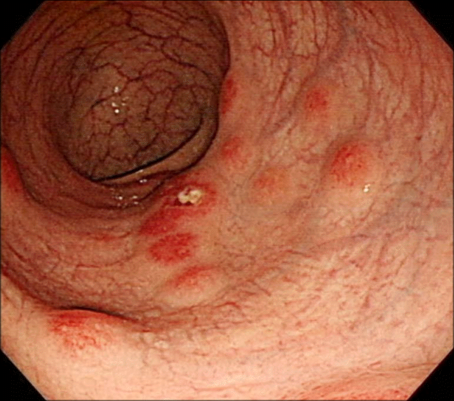
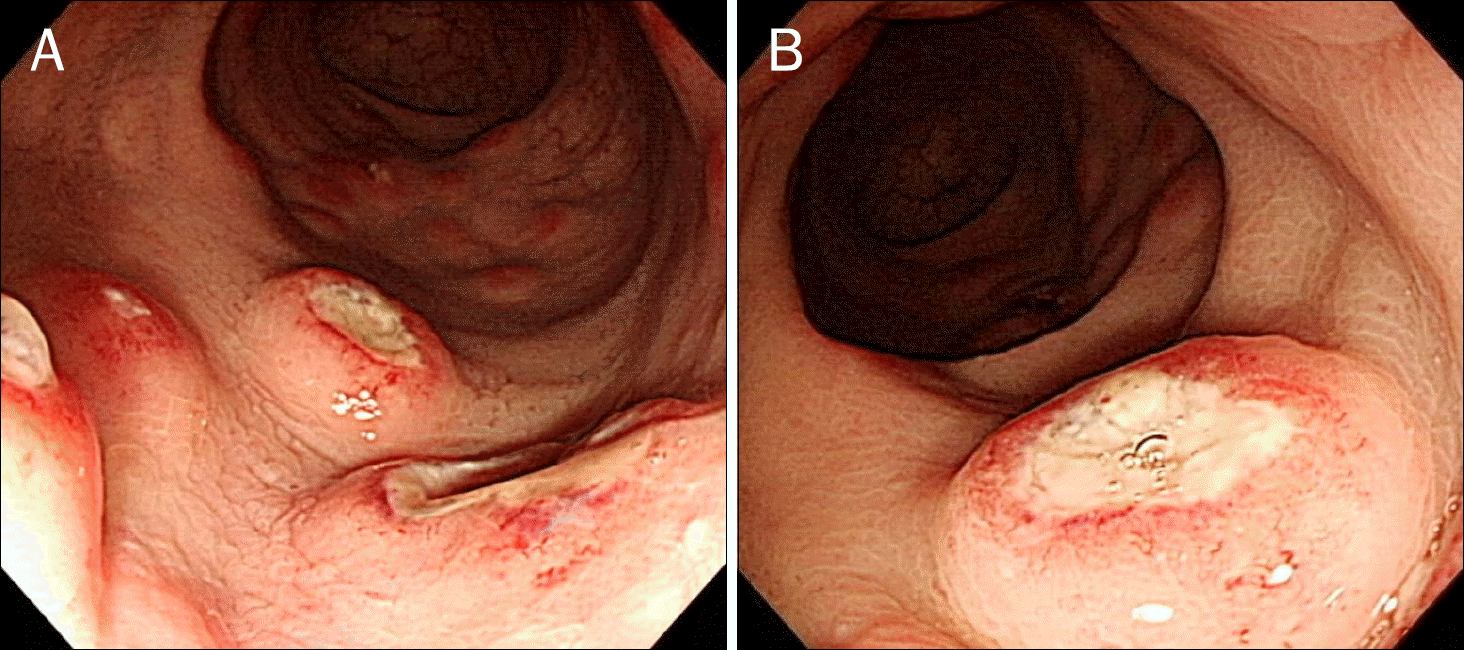
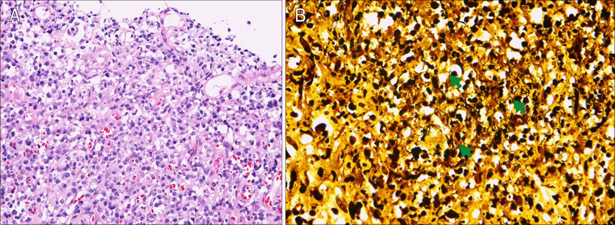
Syphilis is a rare disease in the rectum. It is difficult to diagnose because the characteristics of the rectal syphilis rectal lesion are highly varied. The endoscopic findings of rectal syphilis are proctitis, ulcers, and masses. If rectal syphilis is suspected to be the cause for rectal lesions, it is important for physicians to consider the sexual history and sexual orientation of the patient. We report a case of incidental rectal syphilis in a 41-year-old man diagnosed during a regular medical check-up.
Go to : 
References
1. Choi KC, Song JY. Recent trends in clinical observation of syphilis and consideration for laboratory tests. J Korean Med Assoc. 2009; 52:1100–1106.

2. Gopal P, Shah RB. Primary anal canal syphilis in men: The clinicopathologic spectrum of an easily overlooked diagnosis. Arch Pathol Lab Med. 2015; 139:1156–1160.

3. Korea Centers for Disease Control and Prevention. 2013 White book of disease control. Cheongju: Korea Centers for Disease Control and Prevention;2014.
4. Ghanem KG, Workowski KA. Management of adult syphilis. Clin Infect Dis. 2011; 53(Suppl 3):S110–S128.

5. Kim T. Treatment and management of sexually transmitted diseases. J Korean Med Assoc. 2008; 51:884–896.

6. Song SH, Jang I, Kim BS, et al. A case of primary syphilis in the rectum. J Korean Med Sci. 2005; 20:886–887.

Go to : 


 PDF
PDF ePub
ePub Citation
 Print
Print


 XML Download