References
1. Khera G, Shea AJ, Lambrianides AL. A new approach in the treatment of faecaloma of the colon. Hosp Med. 2005; 66:246–247.

2. Lee JJ, Kim JW. Successful removal of hard sigmoid fecaloma using endoscopic cola injection. Korean J Gastroenterol. 2015; 66:46–49.

3. Kim KH, Kim YS, Seo GS, Choi CS, Choi SC. A case of fecaloma resulting in the rectosigmoid megacolon. Korean J Neurogastroenterol Motil. 2007; 13:81–85.
4. Cid AA, Pietruk T, Bidari CZ, Ehrinpreis MN. Cecal fecaloma mimicking colonic neoplasm. Dig Dis Sci. 1981; 26:1134–1137.

5. Garisto JD, Campillo L, Edwards E, Harbour M, Ermocilla R. Giant fecaloma in a 12-year-old-boy: a case report. Cases J. 2009; 2:127.

6. Lal S, Brown GN. Some unusual complications of fecal impaction. Am J Proctol. 1967; 18:226–231.
8. Yuan R, Zhao GG, Papez S, Cleary JP, Heliotis A. Urethral obstruction and bilateral ureteral hydronephroses secondary to fecal impaction. J Clin Gastroenterol. 2000; 30:314–316.

9. Hoballah JJ, Chalmers RT, Sharp WJ, Stokes JB, Corson JD. Fecal impaction as a cause of acute lower limb ischemia. Am J Gastroenterol. 1995; 90:2055–2057.
10. Schein M, Wittmann DH, Aprahamian CC, Condon RE. The abdominal compartment syndrome: the physiological and clinical consequences of elevated intraabdominal pressure. J Am Coll Surg. 1995; 180:745–753.
Fig. 1.
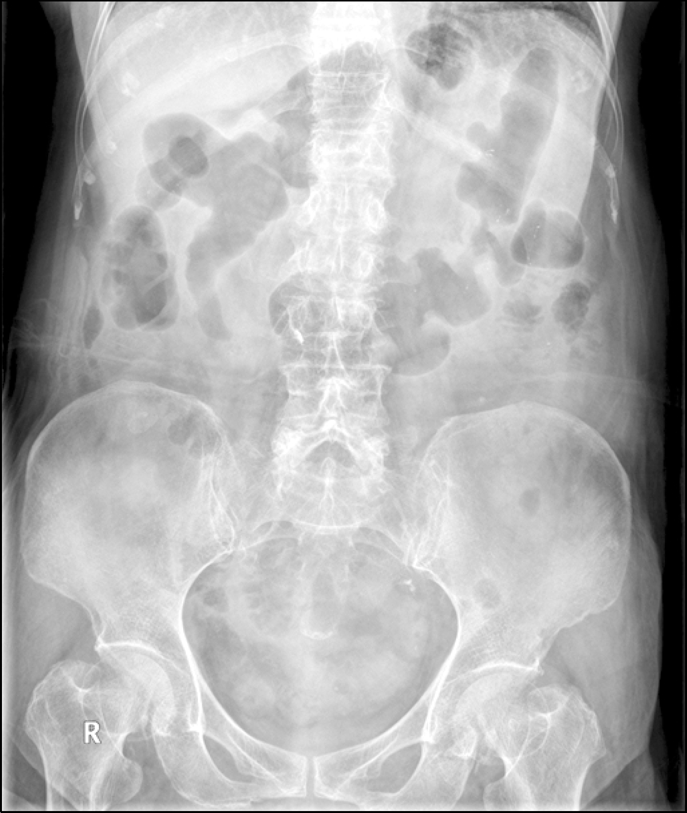
Supine (A) and erect (B) simple abdomen. A fecaloma with calcification is found in the descending colon (arrows), and marked accumulation of bowel gas is present in the transverse and ascending colons.

Fig. 2.
Abdominal CT findings. (A-C) A 4.2 cm sized air-containing fecaloma with calcification is seen in the descending colon (arrows). (D) A large amount of fecal material is noted in the dilated bowel loop of the proximal colon.

Fig. 3.
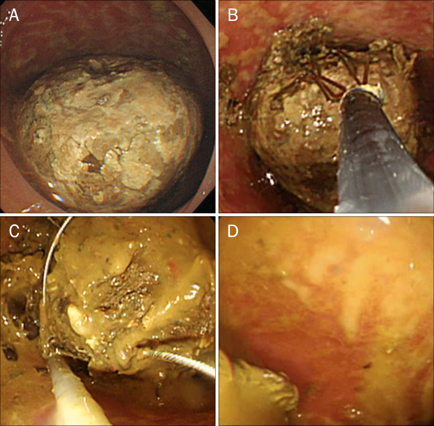
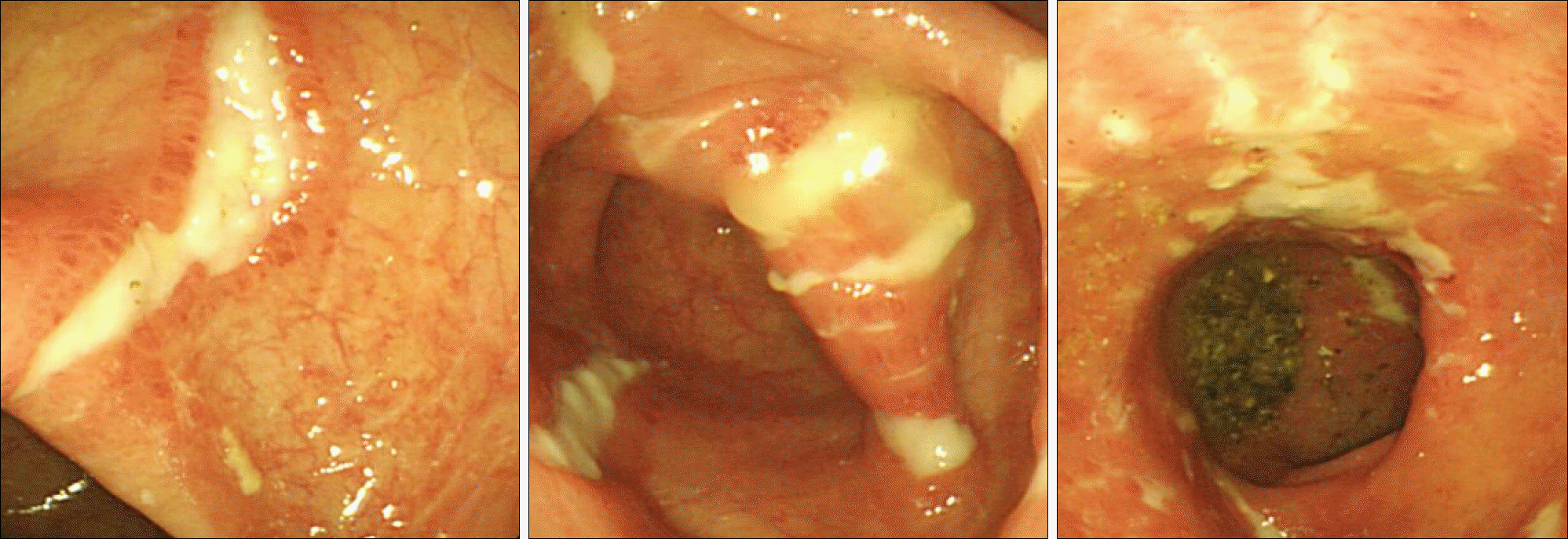
Colonoscopy findings. (A) A large fecaloma is noted at the descending colon. (B, C) Fragmentation of fecaloma with lithotripsy basket and polypectomy snare. (D) After fragmentation, several stercoral ulcers are noted.


 PDF
PDF ePub
ePub Citation
 Print
Print


 XML Download