Abstract
Primary biliary mucosa-associated lymphoid tissue (MALT) lymphoma is extremely rare. We report a case of primary biliary MALT lymphoma with obstructive jaundice diagnosed by endoscopic biopsy, without surgical intervention. Obstructive jaundice was relieved by endoscopic drainage and endoscopic biopsy was done simultaneously during endoscopic retrograde cholangiopancreatography. Unnecessary surgical intervention can be avoided after pathological confirmation of lymphoma. The patient received radiotherapy, and is alive without any evidence of recurrence or biliary obstruction. Diagnosis of primary biliary lymphoma is very difficult because of its low prevalence. However, it should always be considered as a differential diagnosis, since when an accurate diagnosis is made, unnecessary surgical intervention can be avoided.
Go to : 
References
1. Kang HG, Choi JS, Seo JA, et al. A case of primary biliary malignant lymphoma mimicking Klatskin tumor. Korean J Gastroenterol. 2009; 54:191–195.

2. Nguyen GK. Primary extranodal non-Hodgkin's lymphoma of the extrahepatic bile ducts. Report of a case. Cancer. 1982; 50:2218–2222.

3. Kang CS, Lee YS, Kim SM, Kim BK. Primary low-grade B cell lymphoma of mucosa-associated lymphoid tissue type of the common bile duct. J Gastroenterol Hepatol. 2001; 16:949–951.

4. Suzuki S, Tanaka S, Suzuki M, et al. Mucosa-associated lymphoid tissue-type lymphoproliferative lesion of the common bile duct. Hepatogastroenterology. 2004; 51:110–113.
5. Shito M, Kakefuda T, Omori T, Ishii S, Sugiura H. Primary non-Hodgkin's lymphoma of the main hepatic duct junction. J Hepatobiliary Pancreat Surg. 2008; 15:440–443.

6. Yoon MA, Lee JM, Kim SH, et al. Primary biliary lymphoma mimicking cholangiocarcinoma: a characteristic feature of discrep-ant CT and direct cholangiography findings. J Korean Med Sci. 2009; 24:956–959.

7. Cho YH, Byun JH, Kim JH, Lee SS, Kim HJ, Lee MG. Primary malt lymphoma of the common bile duct. Korean J Radiol. 2013; 14:764–768.

8. Boccardo J, Khandelwal A, Ye D, Duke BE. Common bile duct MALT lymphoma: case report and review of the literature. Am Surg. 2006; 72:85–88.

9. Troch M, Kiesewetter B, Raderer M. Recent developments in nongastric mucosa-associated lymphoid tissue lymphoma. Curr Hematol Malig Rep. 2011; 6:216–221.

10. Olszewski AJ, Desai A. Radiation therapy administration and survival in stage I/II extranodal marginal zone B-cell lymphoma of mucosa-associated lymphoid tissue. Int J Radiat Oncol Biol Phys. 2014; 88:642–649.

Go to : 
 | Fig. 1.CT scan of abdomen shows enhancing common bile duct (CBD) wall thickening from (A) intrahepatic bile duct (arrow) to (B) CBD (arrows). (C) Magnetic resonance cholangiopancreatography shows completely separated central main bile ducts and dilatations of intrahepatic bile ducts. Common hepatic duct and CBD duct cannot be seen from hilum to intra-pancreatic portion of CBD (arrows). (D) Cholangiography from the ERCP shows severe CBD stricture (arrows) from hilum to proximal CBD with upstream ductal dilatation. |
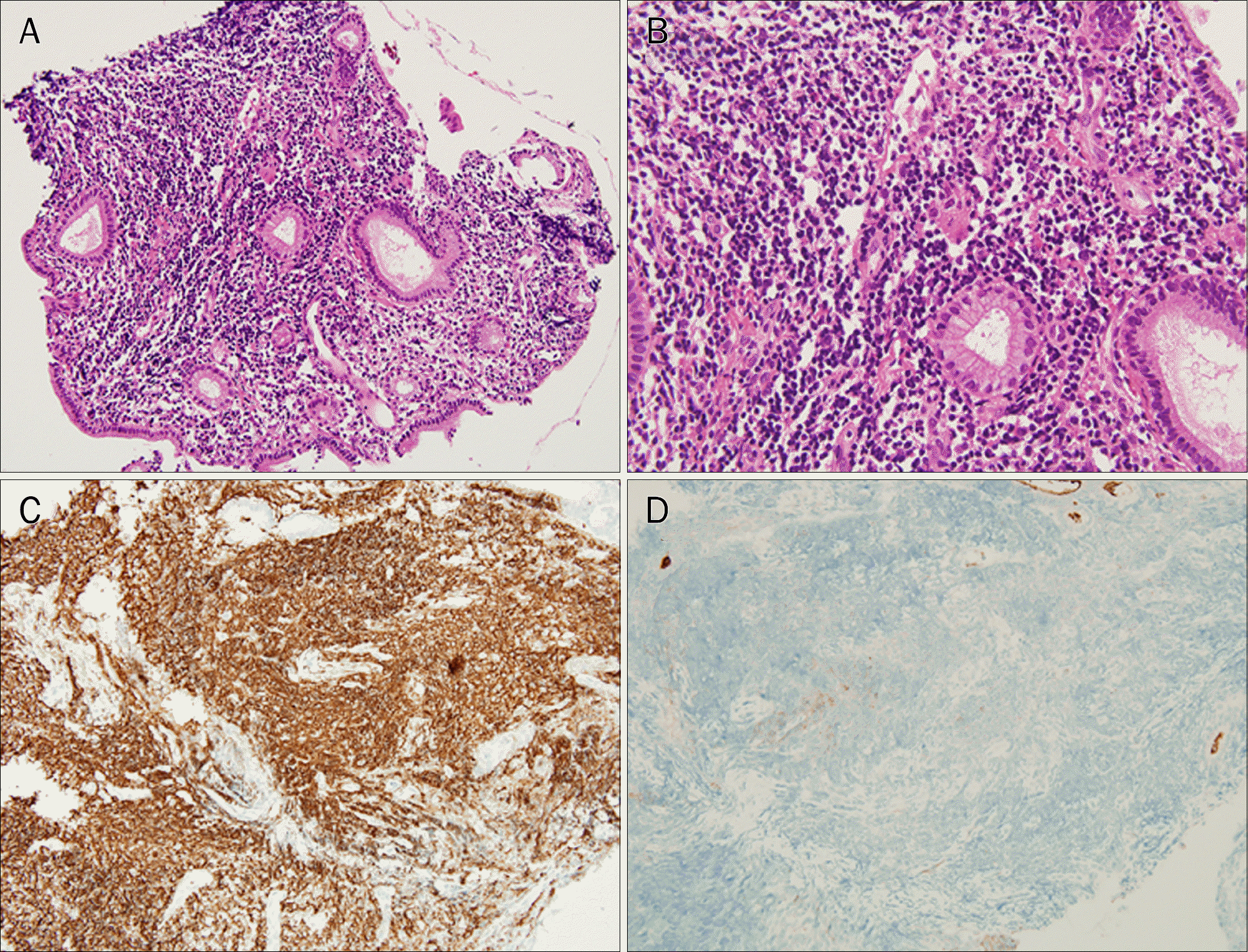
 | Fig. 2.Histopathology and immunohistochemistry showing (A) diffuse infiltration of lymphoid cells (H&E, ×100), (B) B cell proliferation with granulation tissue with lymphoepithelial lesion (H&E, ×200), (C) diffuse immune-positivity for CD20 (×100; monoclonal B cell proliferative lesion) and (D) immune-negativity for CD10 (×100; excluding reactive lymphoid follicle hyperplasia). |
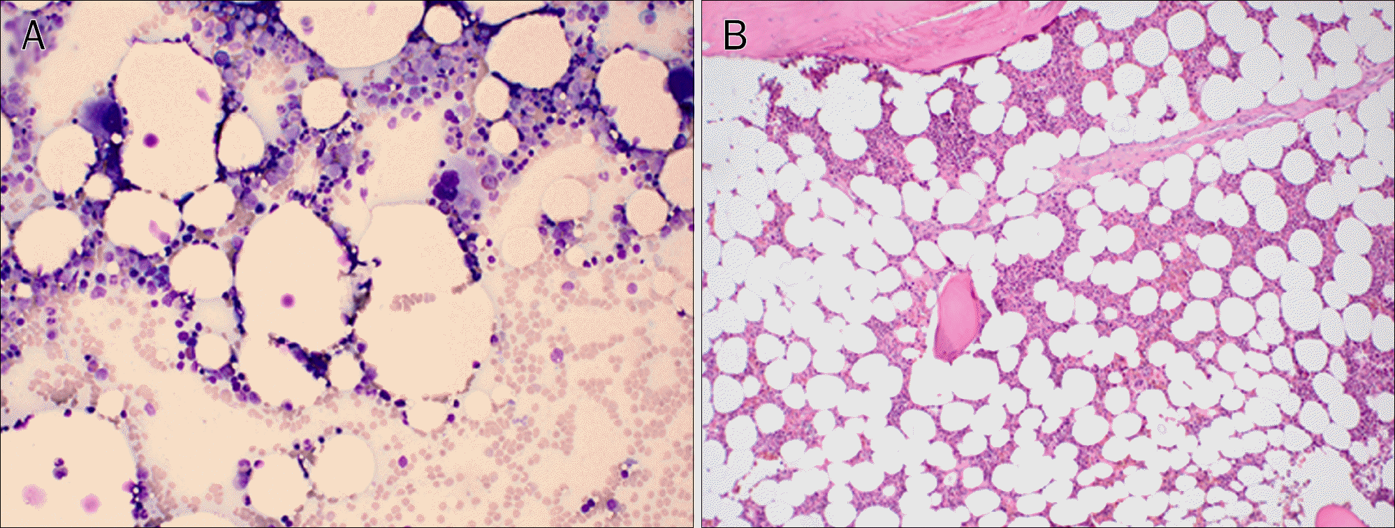
 | Fig. 3.Bone marrow aspiration smear (A; Wright-Giemsa stain, ×200) and biopsy (B; H&E, ×100) show a normo-cellular bone marrow without neoplastic lymphoid cell infiltration. |
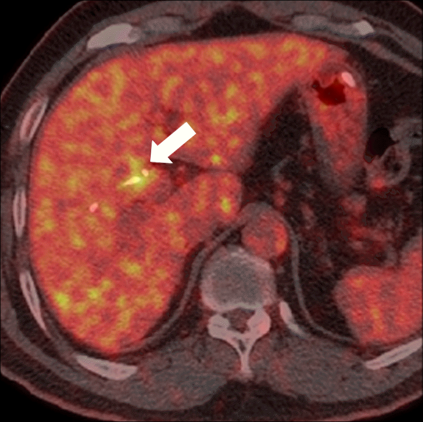
 | Fig. 4.PET shows focal intense F-18 fluorodeoxyglucose uptake (max SUV=6.0) at the hilum of the liver. |
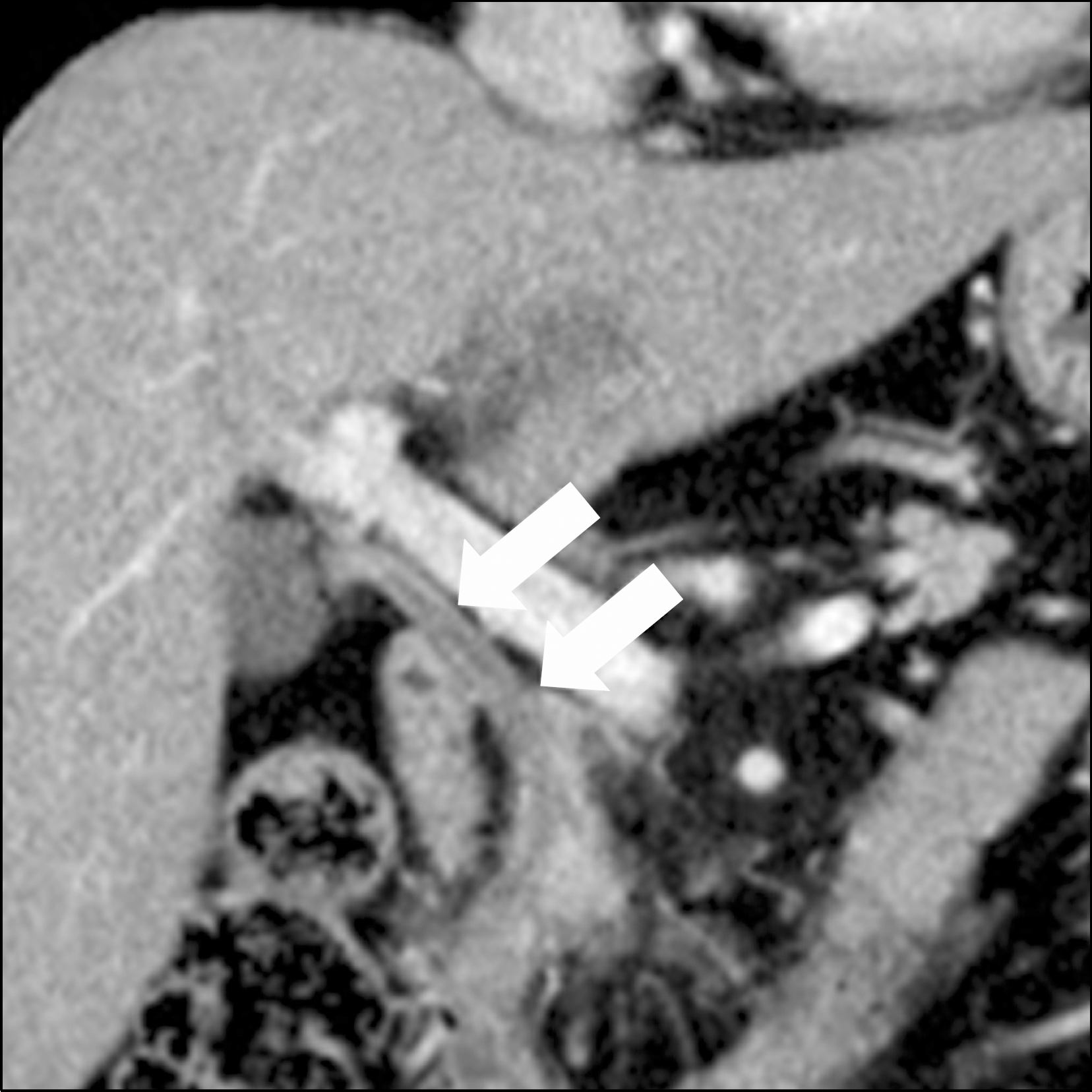
 | Fig. 5.After eight months, CT scan shows improvement of lymphoma involvement in common bile duct with decompressed intrahepatic duct dilatation (arrows). |
Table 1.
Literature Review of Primary Biliary MALT Lymphoma


 PDF
PDF ePub
ePub Citation
 Print
Print


 XML Download