

 PDF
PDF ePub
ePub Citation
 Print
Print


Abstract
Primary hepatic neuroendocrine carcinoma (PHNEC) is rare and its origin is not clearly understood. The coexistence of PHNEC and hepaotcellular carcinoma has been reported in only a few cases. We report a rare case of combined PHNEC and hepaotcellular carcinoma in a patient with liver cirrhosis caused by chronic hepatitis B that resulted in aggressive behavior and poor prognosis.
References
1. Barsky SH, Linnoila I, Triche TJ, Costa J. Hepatocellular carcinoma with carcinoid features. Hum Pathol. 1984; 15:892–894.

2. Artopoulos JG, Destuni C. Primary mixed hepatocellular carcinoma with carcinoid characteristics. A case report. Hepatogastroenterology. 1994; 41:442–444.
3. Tajima Y, Nakajima T, Sugano I, Nagao K, Kondo Y, Saito J. Hepatocellular carcinoma containing endocrine cells. An autopsy report of triplecancer involving the liver, kidney and thyroid. Acta Pathol Jpn. 1992; 42:904–910.
4. Wang JH, Dhillon AP, Sankey EA, Wightman AK, Lewin JF, Scheuer PJ. 'Neuroendocrine' differentiation in primary neoplasms of the liver. J Pathol. 1991; 163:61–67.
5. Zhao M, Laissue JA, Zimmermann A. "Neuroendocrine" differentiation in hepatocellular carcinomas (HCCs): immunohistochemical reactivity is related to distinct tumor cell types, but not to tumor grade. Histol Histopathol. 1993; 8:617–626.
6. Pilichowska M, Kimura N, Ouchi A, Lin H, Mizuno Y, Nagura H. Primary hepatic carcinoid and neuroendocrine carcinoma: clinicopathological and immunohistochemical study of five cases. Pathol Int. 1999; 49:318–324.
7. Kaya G, Pasche C, Osterheld MC, Chaubert P, Fontolliet C. Primary neuroendocrine carcinoma of the liver: an autopsy case. Pathol Int. 2001; 51:874–878.
8. Ishida M, Seki K, Tatsuzawa A, et al. Primary hepatic neuroendocrine carcinoma coexisting with hepatocellular carcinoma in hepatitis C liver cirrhosis: report of a case. Surg Today. 2003; 33:214–218.
9. Yamaguchi R, Nakashima O, Ogata T, Hanada K, Kumabe T, Kojiro M. Hepatocellular carcinoma with an unusual neuroendocrine component. Pathol Int. 2004; 54:861–865.
10. Garcia MT, Bejarano PA, Yssa M, Buitrago E, Livingstone A. Tumor of the liver (hepatocellular and high grade neuroendocrine carcinoma): a case report and review of the literature. Virchows Arch. 2006; 449:376–381.
11. Gould VE, Banner BF, Baerwaldt M. Neuroendocrine neoplasms in unusual primary sites. Diagn Histopathol. 1981; 4:263–277.
12. Hsu W, Deziel DJ, Gould VE, Warren WH, Gooch GT, Staren ED. Neuroendocrine differentiation and prognosis of extrahepatic biliary tract carcinomas. Surgery. 1991; 110:604–610. discussion 610–611.
13. Cheon JH, Park JW, Park KW, et al. The clinical report of 1,078 cases of hepatocellular carcinomas: National Cancer Center experience. Korean J Hepatol. 2004; 10:288–297.
14. Fenoglio LM, Severini S, Ferrigno D, et al. Primary hepatic carcinoid: a case report and literature review. World J Gastroenterol. 2009; 15:2418–2422.
15. Park SH, Kang MJ, Cho JH, et al. Hepatocellular carcinoma with neuroendocrine differentiation: clinical and imaging findings in five patients. J Korean Radiol Soc. 2008; 58:65–71.
16. Bader TR, Semelka RC, Chiu VC, Armao DM, Woosley JT. MRI of carcinoid tumors: spectrum of appearances in the gastrointestinal tract and liver. J Magn Reson Imaging. 2001; 14:261–269.
17. Krohn M, Grieser C, Weichert W, Pascher A, Denecke T. Well-differentiated neuroendocrine carcinoma mimicking an echino-coccus cyst of the liver in CT-MRI findings with hepatocyte specific contrast material. J Gastrointestin Liver Dis. 2011; 20:439–442.
18. Shetty PK, Baliga SV, Balaiah K, Gnana PS. Primary hepatic neuroendocrine tumor: an unusual cystic presentation. Indian J Pathol Microbiol. 2010; 53:760–762.
19. Ramage JK, Ahmed A, Ardill J, et al. Guidelines for the management of gastroenteropancreatic neuroendocrine (including carcinoid) tumours (NETs). Gut. 2012; 61:6–32.
Fig. 1.
Abdominal CT scan showing tumor of S3 with a high density area in the peripheral part of the mass. (A) Arterial phase showed about 2.4 cm hypervascular nodule in S3 of liver. (B) Portal phase showed heterogeneous enhancing mass. (C) Delayed phase showed pseudo washout sign underlying liver cirrhosis.

Fig. 2.
(A) MRI showed peripheral hypervascular mass with pseudo washout appearance and heterogeneous T2 signal. (B) MRI showed restricted diffusion.

Fig. 3.
Cut section of the specimen showed 2.5 cm light gray mass with clear distinction underlying liver cirrhosis.
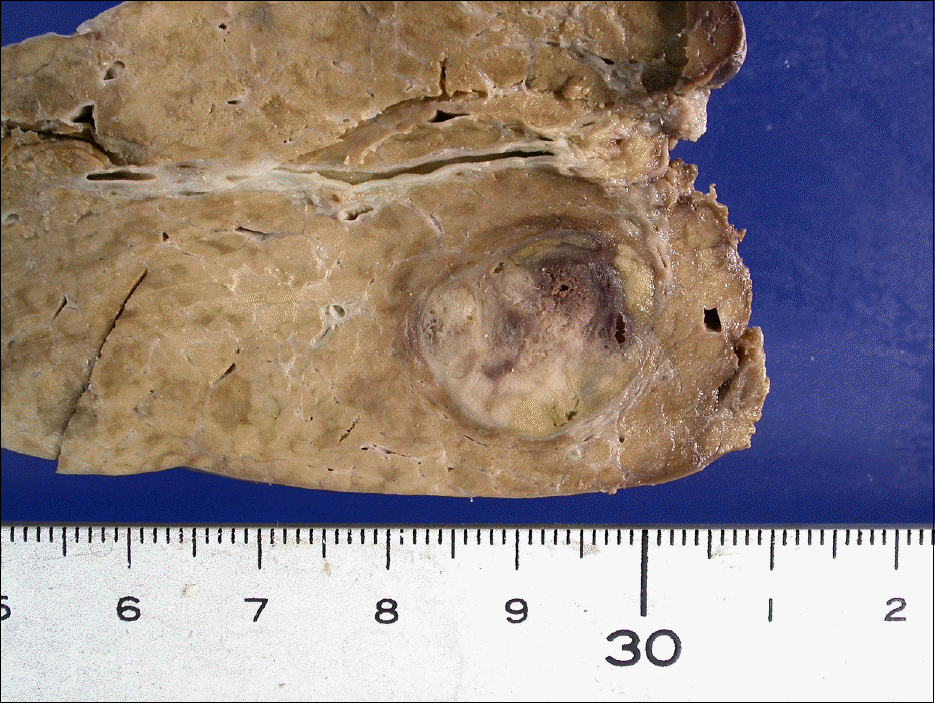
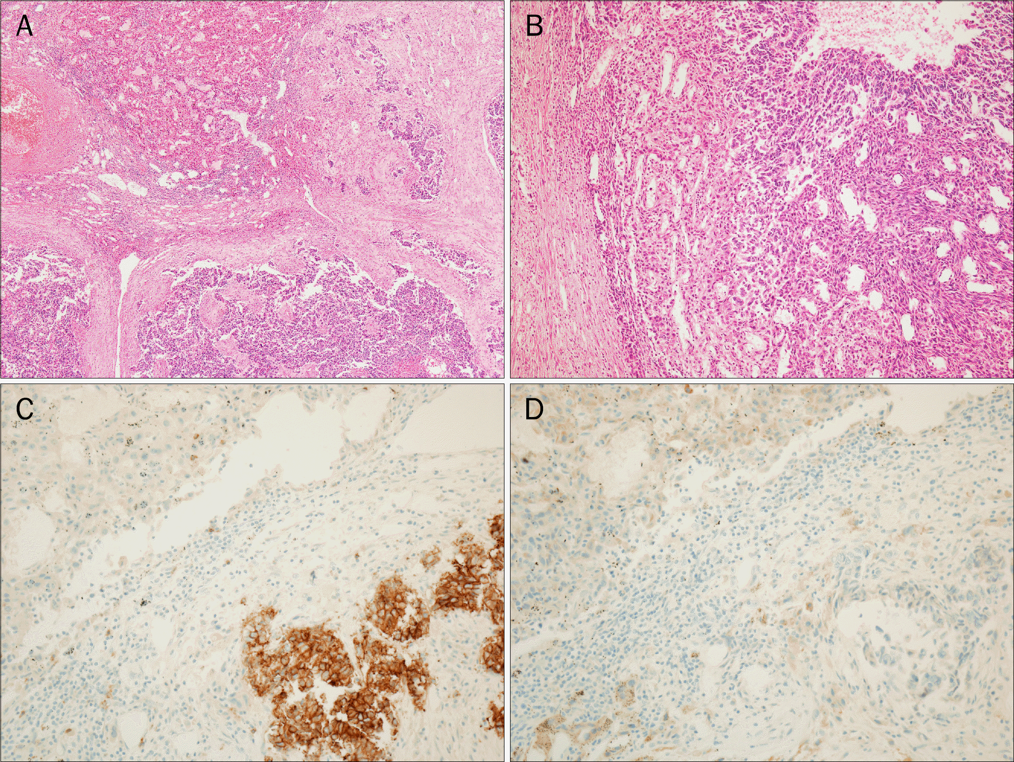
Fig. 4.
(A) The neoplastic cells of the neuroendocrine carcinoma (NEC) are separated from hepatocellular carcinoma (HCC) (H&E, ×40). (B) The transition between HCC (left) and NEC (right) (H&E, ×100). (C) Immunostain for CD56 are strongly positive in small round cells (×200). (D) Immunostain for hepatocyte specific antigen showed positive for HCC (×200).
 XML Download