Abstract
An inverted hyperplastic polyp (IHP) found in stomach is rare and characterized by downward growth of hyperplastic mucosal component into the submucosa. Because of such characteristic, IHP can be misdiagnosed as subepithelial tumor or malignant tumor. In fact, adenocarcinoma was reported to have coexisted with gastric IHP in several previous reports. Because only 18 cases on gastric IHP have been reported in English and Korean literature until now, pathogenesis and clinical features of gastric IHP and correlation with adenocarcinoma have not been clearly established. Herein, we report a case of gastric IHP which was initially misdiagnosed as gastrointestinal stromal tumor and resected using endoscopic submucosal dissection. Literature review of previously published case reports on gastric IHP is also presented.
References
1. Yamashita M, Hirokawa M, Nakasono M, et al. Gastric inverted hyperplastic polyp. Report of four cases and relation to gastritis cystica profunda. APMIS. 2002; 110:717–723.

2. Kamata Y, Kurotaki H, Onodera T, Nishida N. An unusual heterotopia of pyloric glands of the stomach with inverted downgrowth. Acta Pathol Jpn. 1993; 43:192–197.

3. Itoh K, Tsuchigame T, Matsukawa T, Takahashi M, Honma K, Ishimaru Y. Unusual gastric polyp showing submucosal proliferation of glands: case report and literature review. J Gastroenterol. 1998; 33:720–723.

4. Katz LB, Tenembaum MM, Kreel I. Gastric hamartomatous polyps in the absence of familial polyposis: report of two cases. Mt Sinai J Med. 1982; 49:426–429.
5. Hanada M, Takami M, Hirata K, Kishi T, Nakajima T. Hyperplastic fundic gland polyp of the stomach. Acta Pathol Jpn. 1983; 33:1269–1277.

6. Carfagna G, Pilato FP, Bordi C, Barsotti P, Riva C. Solitary polypoid hamartoma of the oxyntic mucosa of the stomach. Pathol Res Pract. 1987; 182:326–330.

7. Aoki M, Yoshida M, Saikawa Y, et al. Diagnosis and treatment of a gastric hamartomatous inverted polyp: report of a case. Surg Today. 2004; 34:532–536.

8. Kono T, Imai Y, Ichihara T, et al. Adenocarcinoma arising in gastric inverted hyperplastic polyp: a case report and review of the literature. Pathol Res Pract. 2007; 203:53–56.

9. Ono S, Kamoshida T, Hiroshima Y, et al. A case of early gastric cancer accompanied by a hamartomatous inverted polyp and successfully managed with endoscopic submucosal dissection. Endoscopy. 2007; 39(Suppl 1):E202.

10. Odashima M, Otaka M, Nanjo H, et al. Hamartomatous inverted polyp successfully treated by endoscopic submucosal dissection. Intern Med. 2008; 47:259–262.

11. Kim HS, Hwang EJ, Jang JY, Lee J, Kim YW. Multifocal adenocarcinomas arising within a gastric inverted hyperplastic polyp. Korean J Pathol. 2012; 46:387–391.

12. Lee SJ, Park JK, Seo HI, et al. A case of gastric inverted hyperplastic polyp found with gastritis cystica profunda and early gastric cancer. Clin Endosc. 2013; 46:568–571.

13. Jung M, Min KW, Ryu YJ. Gastric inverted hyperplasic polyp composed only of pyloric glands: a rare case report and review of the literature. Int J Surg Pathol. 2015; 23:313–316.
14. Choi MS, Jin SY, Kim DW, Lee DW, Park SM. A case of gastric inverted hyperplastic polyp associated with gastritis cystica profunda and early gastric carcinoma. Korean J Pathol. 2007; 41:55–58.
Fig. 1.
Pre-procedural findings. (A) An esophagogastroduodenoscopy shows a submucosal mass with central ulceration in high body, great curvature. (B) Endoscopic ultrasonography shows that the lesion is located in the submucosa and has inhomogeneous hypoechogenecity. (C) A computed tomography shows a 1.9 cm sized lesion in high body, posterior wall (arrow).

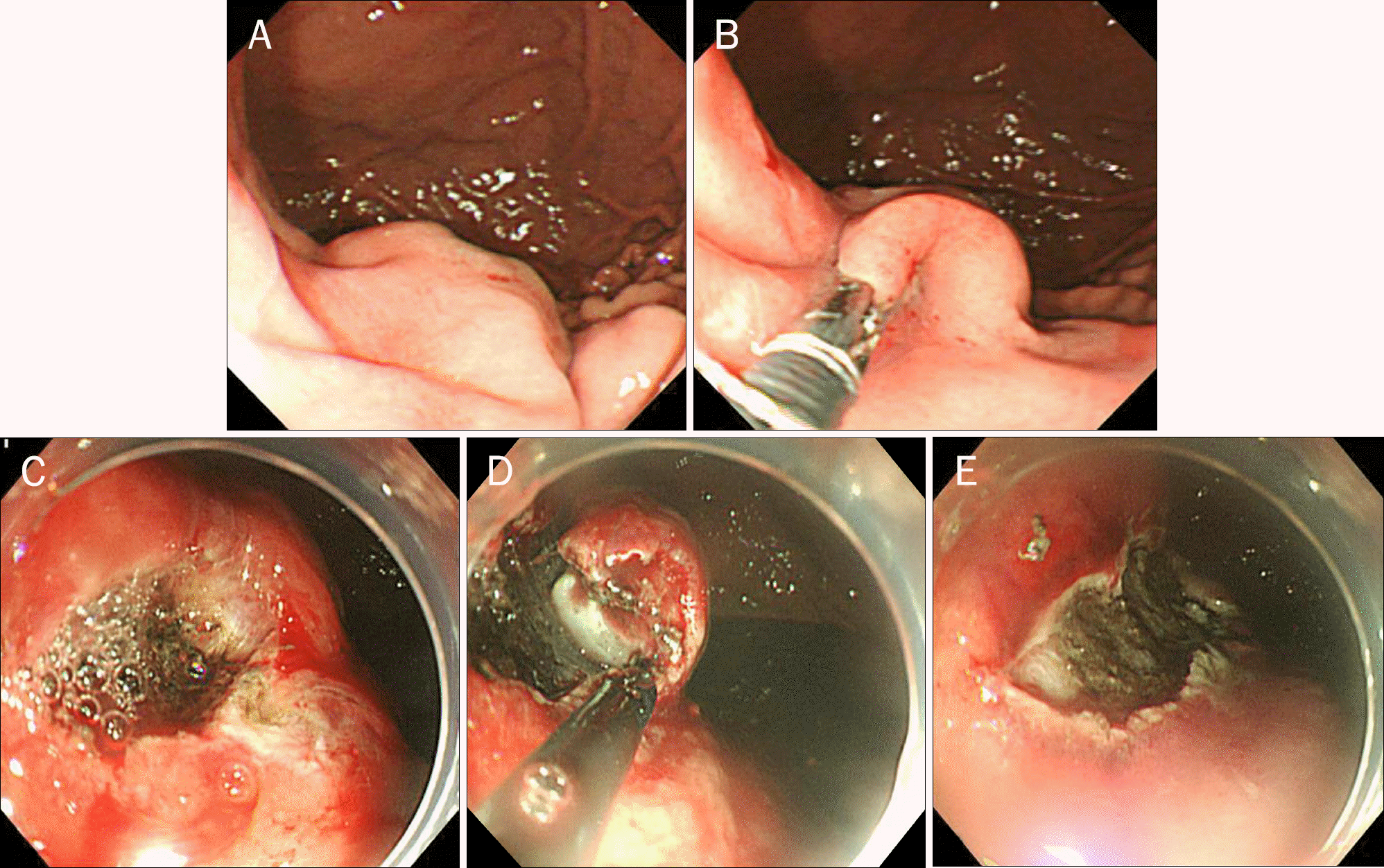
Fig. 2.
Endoscopic submucosal dissection for gastric subepithelial tumor (SET). (A) A large SET is found on the high body, greater curvature side. (B, C) Mucosal layer in the lower part of the tumor is removed using two-channel endoscopy to expose the base of the tumor. (D) Submucosal dissection is performed using insulated tip knife. (E) After resection of the tumor, grossly no remnant tissue is observed.

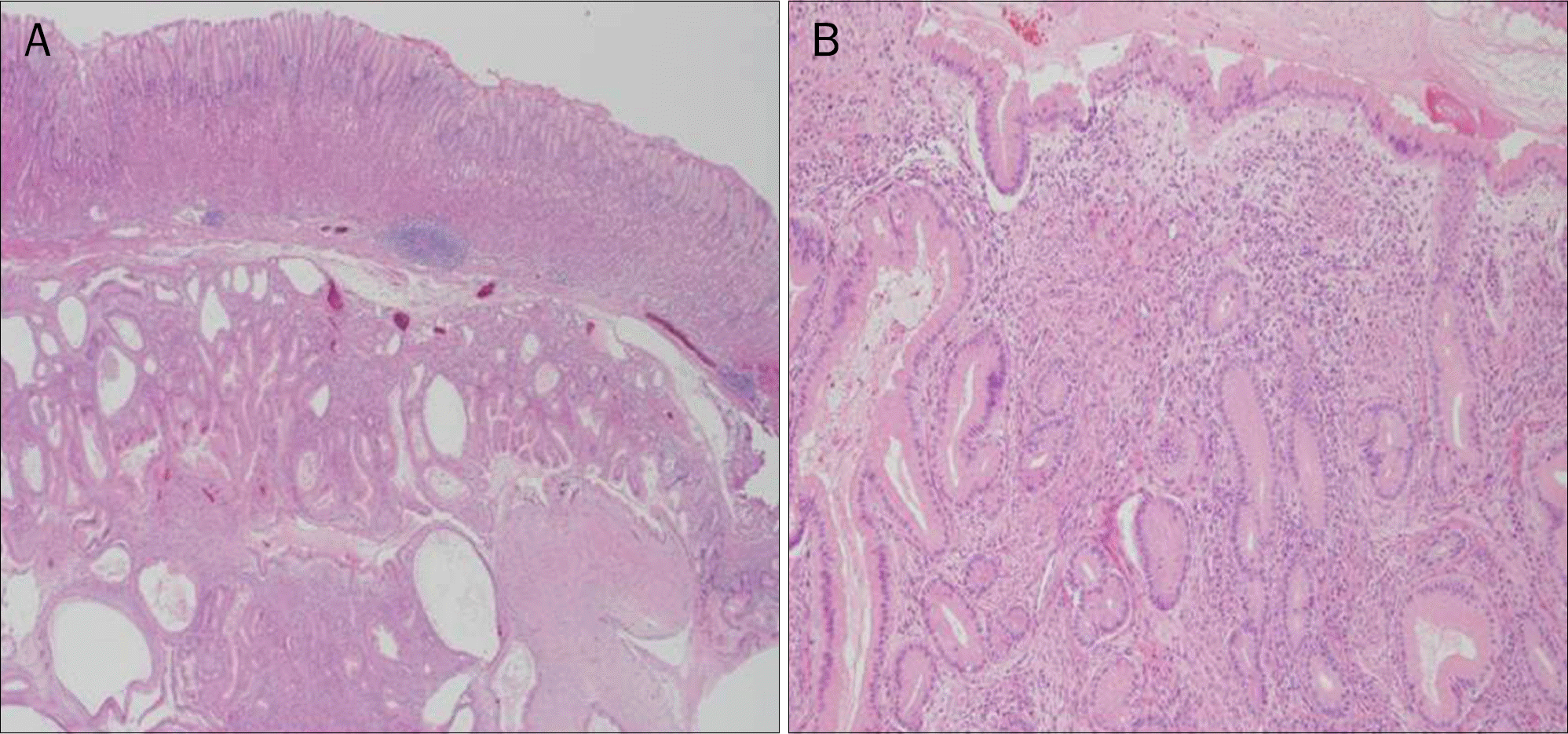
Fig. 3.
Microscopic findings of resected specimen (H&E). (A) Glandular proliferation with cystic dilatation and smooth muscle proliferation are found in submucosa (×12.5). (B) Submucosal lesion consists of foveola-type columnar epithelium (×100).

Table 1.
Case Review of Gastric Inverted Hyperplastic Polyp in English and Korean Literature
| Author | Reported year | Age (yr) | Sex | Symptoms | Location | Gross a | a Size (cm) | Associated findings | Treatment |
|---|---|---|---|---|---|---|---|---|---|
| Yamashita et al.1 | 12002 | 69 | M | Melena | Body | YI | 1×0.9 | AC, GCP | Surgery |
| Yamashita et al.1 | 12002 | 58 | M | Loss of appetite | Cardia | YII | 2.6×2.2 | − | Surgery |
| Yamashita et al.1 | 12002 | 34 | F | Asymptomatic | Body | YIII | 3×3 | − | Surgery |
| Yamashita et al.1 | 12002 | 81 | M | Vomiting, | Fundus | YI | 0.5×0.5 | AC, GCP | Surgery |
| abdominal fullness | |||||||||
| Kamata et al.2 | 1993 | 79 | M | NA | Body | YII | 2.5×1.5 | AC | NA |
| Itoh et al.3 | 1998 | 41 | F | Epigastric discomfort | Fundus | YIV | 2.3×1.8×0.9 | − | EP |
| Katz et al.4 | 1982 | 85 | F | NA | Antrum | NA | 11×5 | NA | NA |
| Katz et al.4 | 1982 | 51 | F | NA | Antrum | NA | 6×4 | NA | NA |
| Hanada et al.5 | 1983 | 47 | F | NA | Fundus | YIV | 1.3×0.9 | NA | NA |
| Carfagna et al.6 | 1987 | 66 | F | NA | Fundus | YIV | 1.5×1.5 | Atrophic gastritis | NA |
| Aoki et al.7 | 2004 | 43 | F | Asymptomatic | Body | YIII | 2.8×2.8 | − | Surgery |
| Kono et al.8 | 2007 | 54 | M | Asymptomatic | Antrum | YIV | 4.5×3.5 | AC, transition zone | Surgery |
| Ono et al.9 | 2007 | 59 | M | Asymptomatic | Body | YIII | NA | AC | ESD |
| Odashima et al.1 | 02008 | 37 | M | Asymptomatic | Fundus | YII | 2.5×2.5 | − | ESD |
| Kim et al.11 | 2012 | 40 | F | Epigastric discomfort | Body | YIII | 3.5×3.2×1.8 | AC, SC, transition zone | ESD |
| Lee et al.12 | 2013 | 77 | M | Asymptomatic | Body | YI | 4.5×3×0.5 | AC, GCP | ESD |
| Jung et al.13 | 2015 | 70 | M | Epigastric discomfort | Body | YI | 1.6×1.5×0.4 | − | ESD |
| Choi et al.14 | 2007 | 71 | M | Epigastric discomfort, | Body | YI | 3.5×3.5 | AC, GCP | Surgery |
| dyspepsia | |||||||||
| Present case | − | 41 | M | Epigastric discomfort | Body | YII | 2×2×1.3 | − | ESD |


 PDF
PDF ePub
ePub Citation
 Print
Print


 XML Download