Abstract
Background/Aims
Although colonoscopy is not indicated in patients with hematochezia, many surgeons, internists, and physicians are recommending colonoscopy for these patients in Korea. The aim of this study is to evaluate the diagnostic value of colonoscopy for patients with hematochezia.
Methods
We retrospectively reviewed the data of colonoscopy between January 2010 and December 2010. A total of 321 patients among 3,038 colonoscopies (10.6%) underwent colonoscopy to evaluate the cause of hematochezia. The patients with previous colorectal surgery (2) or polypectomy (5) were excluded. We analyzed endoscopic diagnoses. Advanced neoplastic polyps were defined as adenomas with villous histology or high grade dysplasia, or adenomas more than 10 mm in diameter.
Results
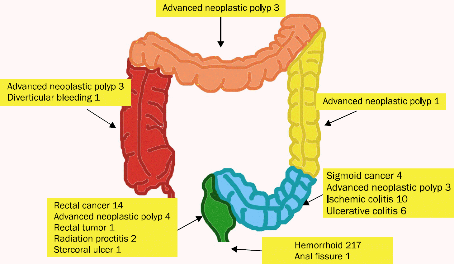
Hemorrhoid was the most common diagnosis (217 cases, 67.6%). Polyps were detected in 93 patients (29.0%), but advanced neoplastic polyps were found in only 14 cases (4.4%). Colorectal cancers were diagnosed in 18 patients (5.6%) including 14 rectal cancers. There was no cancer located above sigmoid-descending junction. Diverticuli were detected in 41 patients (12.8%) but there was only one case of suspected diverticular bleeding. Colitis was diagnosed in 24 patients (7.5%). Other lesions included acute anal fissure, rectal tumor, stercoral ulcer, and radiation proctitis.
Go to : 
References
1. Davila RE, Rajan E, Adler DG, et al. Standards of Practice Committee. ASGE Guideline: the role of endoscopy in the patient with lower-GI bleeding. Gastrointest Endosc. 2005; 62:656–660.

2. Eisen GM, Dominitz JA, Faigel DO, et al. American Society for Gastrointestinal Endoscopy Standards of Practice Committee. Endoscopic therapy of anorectal disorders. Gastrointest Endosc. 2001; 53:867–870.

3. Peytremann-Bridevaux I, Arditi C, Froehlich F, et al. EPAGE II Study Group. Appropriateness of colonoscopy in Europe (EPAGE II). Iron-deficiency anemia and hematochezia. Endoscopy. 2009; 41:227–233.
4. Savides TJ, Jensen DM. Gastrointestinal bleeding. Feldman M, Friedman LS, Brandt LJ, editors. Sleisenger and fordtran's gastrointestinal and liver disease: pathophysiology/diagnosis/management. 10th ed.Philadelphia, PA: Saunders/Elsevier;2016. p. 297–335.e210.

5. Tong GX, Chai J, Cheng J, et al. Diagnostic value of rectal bleeding in predicting colorectal cancer: a systematic review. Asian Pac J Cancer Prev. 2014; 15:1015–1021.

6. Bjerregaard NC, Tøttrup A, Sørensen HT, Laurberg S. Diagnostic value of self-reported symptoms in Danish outpatients referred with symptoms consistent with colorectal cancer. Colorectal Dis. 2007; 9:443–451.

7. Nikpour S, Ali Asgari A. Colonoscopic evaluation of minimal rectal bleeding in average-risk patients for colorectal cancer. World J Gastroenterol. 2008; 14:6536–6540.

8. Korkis AM, McDougall CJ. Rectal bleeding in patients less than 50 years of age. Dig Dis Sci. 1995; 40:1520–1523.

9. Spinzi G, Fante MD, Masci E, et al. SIED Lombardia Working Group, Italy. Lack of colonic neoplastic lesions in patients under 50 yr of age with hematochezia: a multicenter prospective study. Am J Gastroenterol. 2007; 102:2011–2015.

10. Wong RF, Khosla R, Moore JH, Kuwada SK. Consider colonoscopy for young patients with hematochezia. J Fam Pract. 2004; 53:879–884.
11. Bae T, Ha Y, Kim C, et al. Distribution of the colonoscopic adenoma detection rate according to age: is recommending colonoscopy screening for Koreans over the age of 50 safe? Ann Coloproctol. 2015; 31:46–51.

12. Chun JH, Son HJ, Rhee PL, et al. Clinical characteristics of lower gastrointestinal bleeding. Korean J Gastrointest Endosc. 1999; 19:911–917.
13. Noer RJ, Robb HJ, Jacobson LF. Circulatory disturbances produced by acute intestinal distention in the living animal. AMA Arch Surg. 1951; 63:520–528.

Go to : 
 | Fig. 1.Location of clinically significant lesions causing hematochezia. The most significant lesions were located in the sigmoid colon and rectum. |
Table 1.
Causes of Hematochezia according to Age Group


 PDF
PDF ePub
ePub Citation
 Print
Print


 XML Download