Abstract
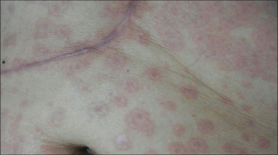
Sorafenib is currently the only targeted therapy available for advanced stage hepatocellular carcinoma (HCC). Cutaneous adverse events associated with sorafenib treatment include hand-foot skin reaction, but there has been no report of drug reaction (or rash) with eosinophilia and systemic symptoms (DRESS) syndrome. Here, we report a case of 72-year-old man with HCC and alcoholic liver cirrhosis who developed skin eruptions, fever, eosinophilia, and deteriorated hepatic and renal function under sorafenib treatment. He has since successfully recovered with conservative care.
Go to : 
References
1. Llovet JM, Ricci S, Mazzaferro V, et al. SHARP Investigators Study Group. Sorafenib in advanced hepatocellular carcinoma. N Engl J Med. 2008; 359:378–390.

2. Bruix J, Raoul JL, Sherman M, et al. Efficacy and safety of sorafenib in patients with advanced hepatocellular carcinoma: sub-analyses of a phase III trial. J Hepatol. 2012; 57:821–829.

3. Sohn KH, Oh SY, Lim KW, Kim MY, Lee SY, Kang HR. Sorafenib induces delayed-onset cutaneous hypersensitivity: a case series. Allergy Asthma Immunol Res. 2015; 7:304–307.

5. Autier J, Escudier B, Wechsler J, Spatz A, Robert C. Prospective study of the cutaneous adverse effects of sorafenib, a novel multikinase inhibitor. Arch Dermatol. 2008; 144:886–892.

6. Cohen PR. Sorafenib-associated facial acneiform eruption. Dermatol Ther (Heidelb). 2015; 5:77–86.

7. Namba M, Tsunemi Y, Kawashima M. Sorafenib-induced erythema multiforme: three cases. Eur J Dermatol. 2011; 21:10151016.

8. Ikeda M, Fujita T, Amoh Y, Mii S, Matsumoto K, Iwamura M. Stevens-Johnson syndrome induced by sorafenib for metastatic renal cell carcinoma. Urol Int. 2013; 91:482–483.

9. Bouvresse S, Valeyrie-Allanore L, Ortonne N, et al. Toxic epidermal necrolysis, DRESS, AGEP: do overlap cases exist? Orphanet J Rare Dis. 2012; 7:72.

10. Roujeau JC, Stern RS. Severe adverse cutaneous reactions to drugs. N Engl J Med. 1994; 331:1272–1285.

11. Choudhary S, McLeod M, Torchia D, Romanelli P. Drug reaction with eosinophilia and systemic symptoms (DRESS) syndrome. J Clin Aesthet Dermatol. 2013; 6:31–37.
12. Eshki M, Allanore L, Musette P, et al. Twelve-year analysis of severe cases of drug reaction with eosinophilia and systemic symptoms: a cause of unpredictable multiorgan failure. Arch Dermatol. 2009; 145:67–72.

13. Peyrière H, Dereure O, Breton H, et al. Network of the French Pharmacovigilance Centers. Variability in the clinical pattern of cutaneous side-effects of drugs with systemic symptoms: does a DRESS syndrome really exist? Br J Dermatol. 2006; 155:422–428.

14. Kardaun SH, Sidoroff A, Valeyrie-Allanore L, et al. Variability in the clinical pattern of cutaneous side-effects of drugs with systemic symptoms: does a DRESS syndrome really exist? Br J Dermatol. 2007; 156:609–611.

15. Wongkitisophon P, Chanprapaph K, Rattanakaemakorn P, Vachiramon V. Six-year retrospective review of drug reaction with eosinophilia and systemic symptoms. Acta Derm Venereol. 2012; 92:200–205.

16. Cacoub P, Musette P, Descamps V, et al. The DRESS syndrome: a literature review. Am J Med. 2011; 124:588–597.

17. Fernando SL. Drug-reaction eosinophilia and systemic symptoms and drug-induced hypersensitivity syndrome. Australas J Dermatol. 2014; 55:15–23.

18. Kirchhof MG, Miliszewski MA, Sikora S, Papp A, Dutz JP. Retrospective review of Stevens-Johnson syndrome/toxic epidermal necrolysis treatment comparing intravenous immunoglobulin with cyclosporine. J Am Acad Dermatol. 2014; 71:941–947.

19. Koštál M, Bláha M, Lánská M, et al. Beneficial effect of plasma exchange in the treatment of toxic epidermal necrolysis: a series of four cases. J Clin Apher. 2012; 27:215–220.
Go to : 
 | Fig. 1.Abdominal skin lesion at admission. Multiple targetoid and patch-like rash occurred in the lateral part of right thigh and spread to whole body. |
Table 1.
Serial Laboratory Results


 PDF
PDF ePub
ePub Citation
 Print
Print


 XML Download