Abstract
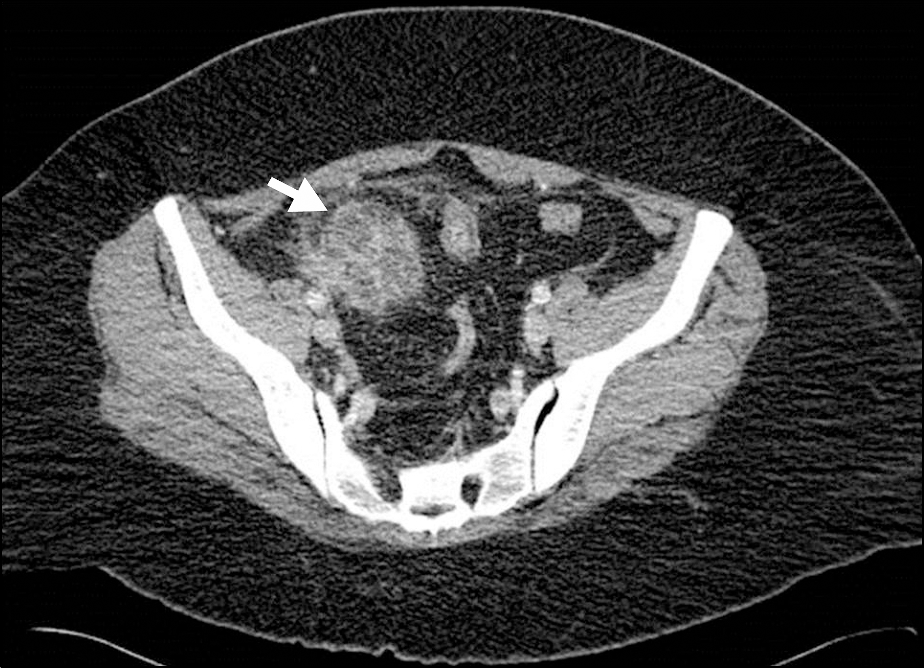
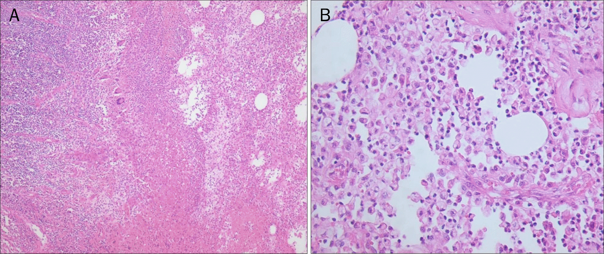
Xanthogranulomatous inflammation is an acute or chronic inflammatory condition most frequently reported in pyelonephritis and cholecystitis. However, the involvement of the terminal ileum is extremely rare. Its clinical significance is that it can mimic a malignant lesion clinically and intraoperatively, as well as radiographically. A 34-year-old European ethnic female presented with gradually aggravated abdominal pain in right lower quadrant for 15 days. There was no significant medical, surgical or traumatic history, except class III obesity (BMI, 41.0 kg/m2). An abdominal CT showed about a 4.7×3.7 cm sized, mass-like lesion in the terminal ileum. Despite symptomatic treatment, her clinical symptoms did not improve. After six days, she underwent a laparoscopic ileocecectomy. Pathologic findings showed extensive inflammation with occasional multinucleated giant cells and aggregates of foamy histiocytes, consistent with xanthogranulomatous inflammation. Here, we present a case of xanthogranulomatous inflammation in the terminal ileum presenting as subacute abdominal pain and a mass on imaging study. Xanthogranulomatous inflammation should be added to the differential diagnosis of patients with a suspected mass-like lesion in the terminal ileum.
Go to : 
References
1. Addario Chieco P, Antolino L, Giaccaglia V, et al. Acute abdomen: rare and unusual presentation of right colic xanthogranulo-matosis. World J Gastroenterol. 2014; 20:8717–8721.
2. Cozzutto C, Carbone A. The xanthogranulomatous process. Xanthogranulomatous inflammation. Pathol Res Pract. 1988; 183:395–402.
3. Anadol AZ, Gonul II, Tezel E. Xanthogranulomatous inflammation of the colon: a rare cause of cecal mass with bleeding. South Med J. 2009; 102:196–199.
4. Oh YH, Seong SS, Jang KS, et al. Xanthogranulomatous inflammation presenting as a submucosal mass of the sigmoid colon. Pathol Int. 2005; 55:440–444.

5. Yoon JS, Jeon YC, Kim TY, et al. Xanthogranulomatous inflammation in terminal ileum presenting as an appendiceal mass: case report and review of the literature. Clin Endosc. 2013; 46:193–196.

6. Wong KC, Tsui WM, Chang SJ. Xanthogranulomatous inflammation of terminal ileum: report of a case with small bowel involvement. Hong Kong Med J. 2015; 21:69–72.

7. Bailey MB, Beck SJ. Xanthogranulomatous inflammation of the sigmoid and small bowel presenting as Crohn's disease. Am Surg. 2014; 80:E38–E39.

8. Osterlind S. Über pyelonephritis xanthomatosa. Acta Chir Scand. 1944; 90:369–376.
9. Son J, Raetskaya-Solntseva O, Tirman PA, Waters GS, Kelly MG. Xanthogranulomatous oophoritis presenting as an adnexal mass and bowel obstruction: a case report. J Reprod Med. 2015; 60:273–276.
10. Nishimura M, Nishihira T, Hirose T, et al. Xanthogranulomatous pancreatitis mimicking a malignant cystic tumor of the pancreas: report of a case. Surg Today. 2011; 41:1310–1313.

11. Tam VK, Kung WH, Li R, Chan KW. Renal parenchymal mala-coplakia: a rare cause of ARF with a review of recent literature. Am J Kidney Dis. 2003; 41:E13–E17.

12. Keitel E, Pêgas KL, do Nascimento Bittar AE, dos Santos AF, da Cas Porto F, Cambruzzi E. Diffuse parenchymal form of malako-plakia in renal transplant recipient: a case report. Clin Nephrol. 2014; 81:435–439.

13. Sumer Y, Yilmaz B, Emre B, Ugur C. Abdominal mass secondary to actinomyces infection: an unusual presentation and its treatment. J Postgrad Med. 2004; 50:115–117.
14. Kim KH, Lee J, Cho HJ, et al. A case of abdominal wall actinomycosis. Korean J Gastroenterol. 2015; 65:236–240.

15. Shalev E, Zuckerman H, Rizescu I. Pelvic inflammatory pseudotumor (xanthogranuloma). Acta Obstet Gynecol Scand. 1982; 61:285–286.

16. Wen H, Gris D, Lei Y, et al. Fatty acid-induced NLRP3-ASC inflammasome activation interferes with insulin signaling. Nat Immunol. 2011; 12:408–415.

17. Jung SH, Kim JH. Cecal cancer with xanthogranulomatous inflammation. J Korean Surg Soc. 2008; 74:392–395.
Go to : 
 | Fig. 1.CT scan of abdomen. Seen is an approximately 4.7×3.7 cm sized, mass-like lesion (white arrow) in the terminal ileum. |
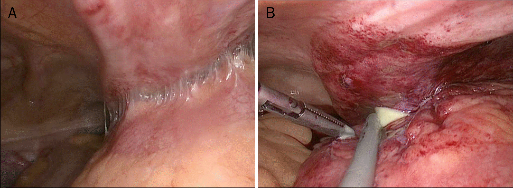
 | Fig. 2.Intraoperative laparoscopic findings. (A) Inflammation and phlegmon with adhesions between the terminal ileum and abdominal wall.(B) Abscess pocket with pus discharges of the terminal ileum. |
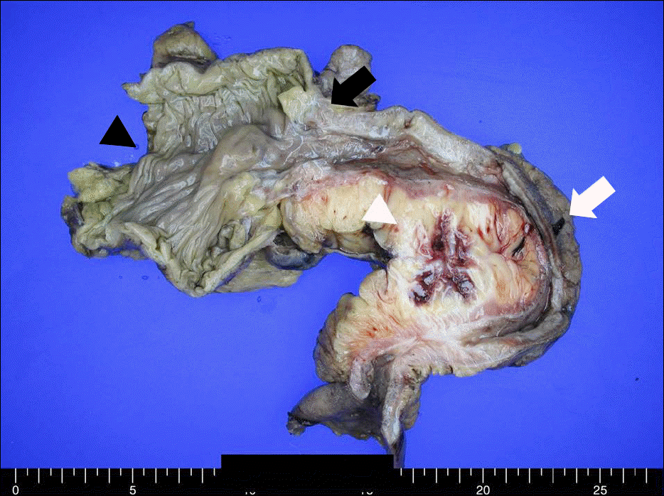
 | Fig. 3.Gross finding of resected ileocecal segment. It revealed mass-like thickening of the mesenteric fat with hemorrhage and adhesion at the terminal ileum. White arrow, terminal ileum; white arrowhead, mass-like lesion; black arrow, ileocecal valve; black arrowhead, cecum. |
 | Fig. 4.Microscopic findings (H&E) of the resected specimen (A) showed extensive inflammation with occasional multinucleated giant cells and fibrosis (×100). (B) Higher magnification revealed aggregates of foamy histiocytes (×400). |
Table 1.
Review of Reported Cases of Small Bowel Involvement in Xanthogranulomatous Inflammation (XGI)


 PDF
PDF ePub
ePub Citation
 Print
Print


 XML Download