Abstract
Pneumatosis cystoides intestinalis (PCI) is a rare condition characterized by multiple gas-filled cysts of varying size in the wall of gastrointestinal tract. PCI may idiopathic or secondary to various disorders. The etiology and pathogenesis of PCI are unclear. Treatment is usually conservative, and includes oxygen and antibiotics therapy. Surgery is reserved for cases of suspected inconvertible intestinal obstruction or perforation. Eleven patients who were diagnosed with PI between 2005 and 2015 were reviewed. We report three cases of PCI and describe causes and complications. The most important point in the treatment of PCI is to determine whether the patient needs surgery. Conservative care should be considered first if the patient is stable. If any complication is observed, such as ischemia in the intestine, surgery is needed. It is important to choose the best treatment based on prognostic factors and CT findings.
Go to : 
References
1. Szucs RA, Wolf EL, Gramm HF, Scholz FJ, Eisenberg RL, Hall DA. Miscellaneous abnormalities of the colon. Gore RM, Levine MS, editors. Textbook of gastrointestinal radiology. 2nd ed.Philadelphia: W.B. Saunders;2000. p. 1114–1115.
2. Höer J, Truong S, Virnich N, Füzesi L, Schumpelick V. Pneumatosis cystoides intestinalis: confirmation of diagnosis by endoscopic puncture a review of pathogenesis, associated disease and therapy and a new theory of cyst formation. Endoscopy. 1998; 30:793–799.

3. Han BG, Lee JM, Yang JW, Kim MS, Choi SO. Pneumatosis intestinalis associated with immune-suppressive agents in a case of minimal change disease. Yonsei Med J. 2002; 43:686–689.

4. Morris MS, Gee AC, Cho SD, et al. Management and outcome of pneumatosis intestinalis. Am J Surg. 2008; 195:679–682. discussion 682–673.

5. Kim HL, Lee HK, Park SJ, et al. Pneumatosis intestinalis: CT findings and clinical features. J Korean Radiol Soc. 2008; 58:149–154.

6. Heng Y, Schuffler MD, Haggitt RC, Rohrmann CA. Pneumatosis intestinalis: a review. Am J Gastroenterol. 1995; 90:1747–1758.
7. Jamart J. Pneumatosis cystoides intestinalis. A statistical study of 919 cases. Acta Hepatogastroenterol (Stuttg). 1979; 26:419–422.
8. Nelson SW. Extraluminal gas collections due to diseases of the gastrointestinal tract. Am J Roentgenol Radium Ther Nucl Med. 1972; 115:225–248.

10. Caudill JL, Rose BS. The role of computed tomography in the evaluation of pneumatosis intestinalis. J Clin Gastroenterol. 1987; 9:223–226.

11. Ham JH, Kim TH, Han SW, et al. A case of pneumatosis cystoides intestinalis: diagnosed by CT colonoscopy. Korean J Gastroenterol. 2007; 50:334–339.
12. Wiesner W, Mortelé KJ, Glickman JN, Ji H, Ros PR. Pneumatosis intestinalis and portomesenteric venous gas in intestinal ischemia: correlation of CT findings with severity of ischemia and clinical outcome. AJR Am J Roentgenol. 2001; 177:1319–1323.
Go to : 
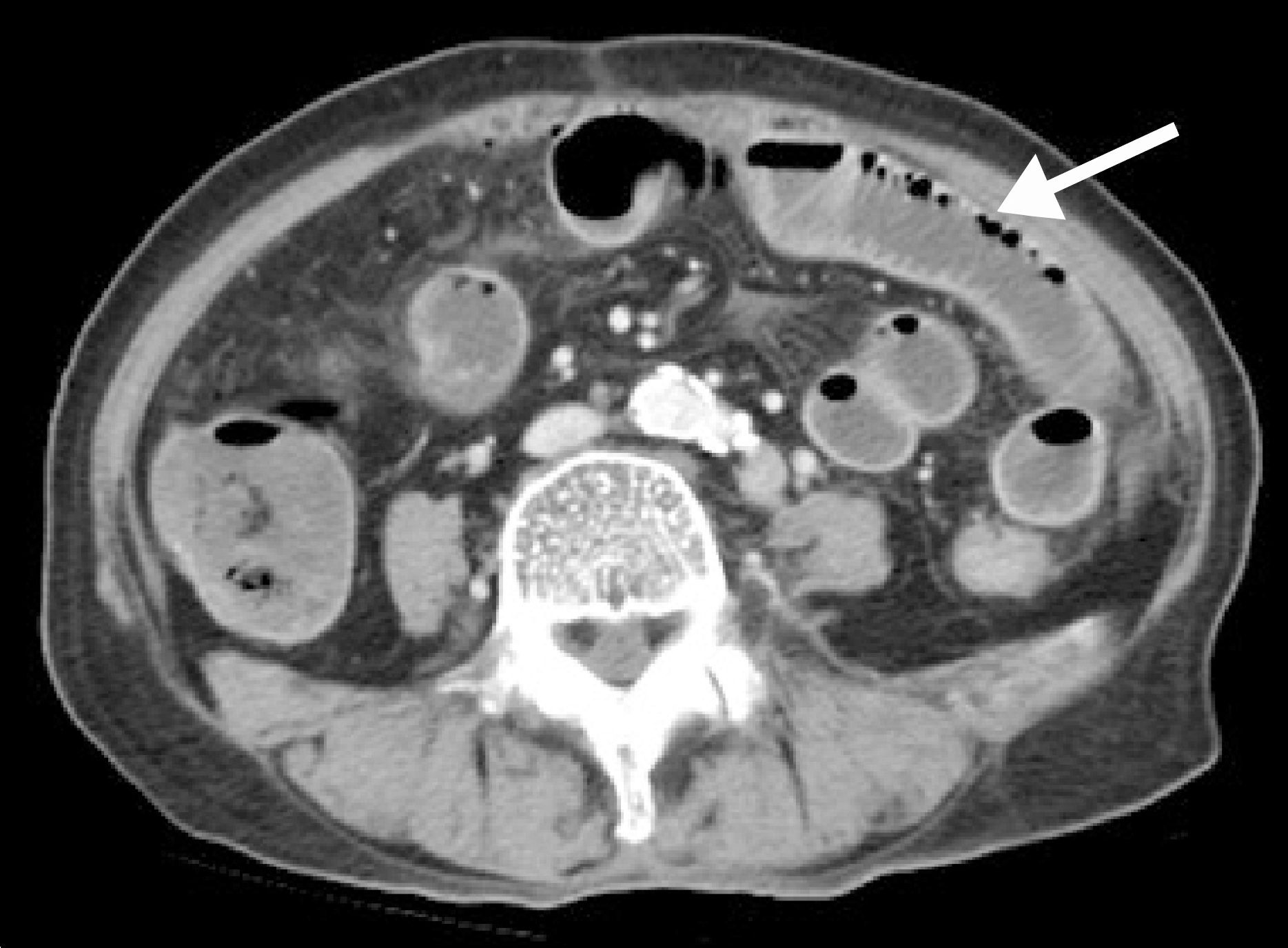
 | Fig. 1.Contrast enhanced CT scan shows intramural bubble gas-like pneumatosis intestinalis (arrow) in the small bowel (case 1). |
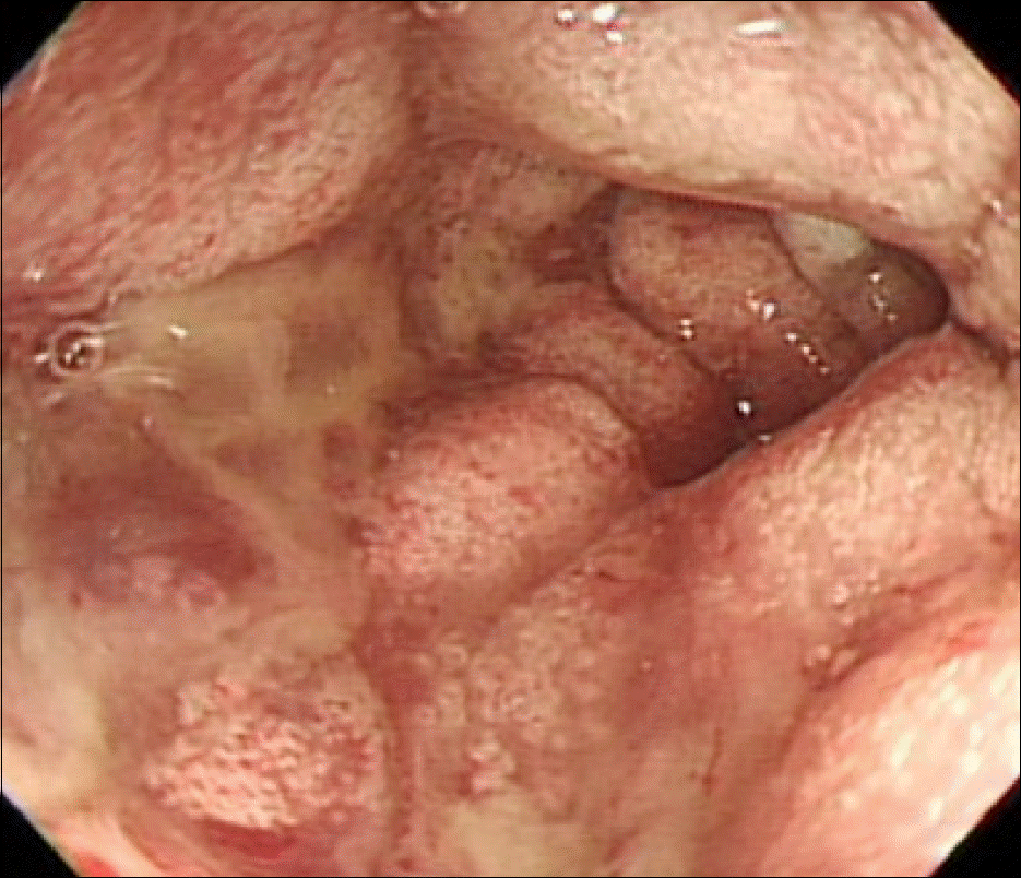
 | Fig. 2.Colonoscopic findings show several polypoid mucosal elevations of variable size, and demarcated erythematous friable mucosal changes at distal descending colon (case 1). |
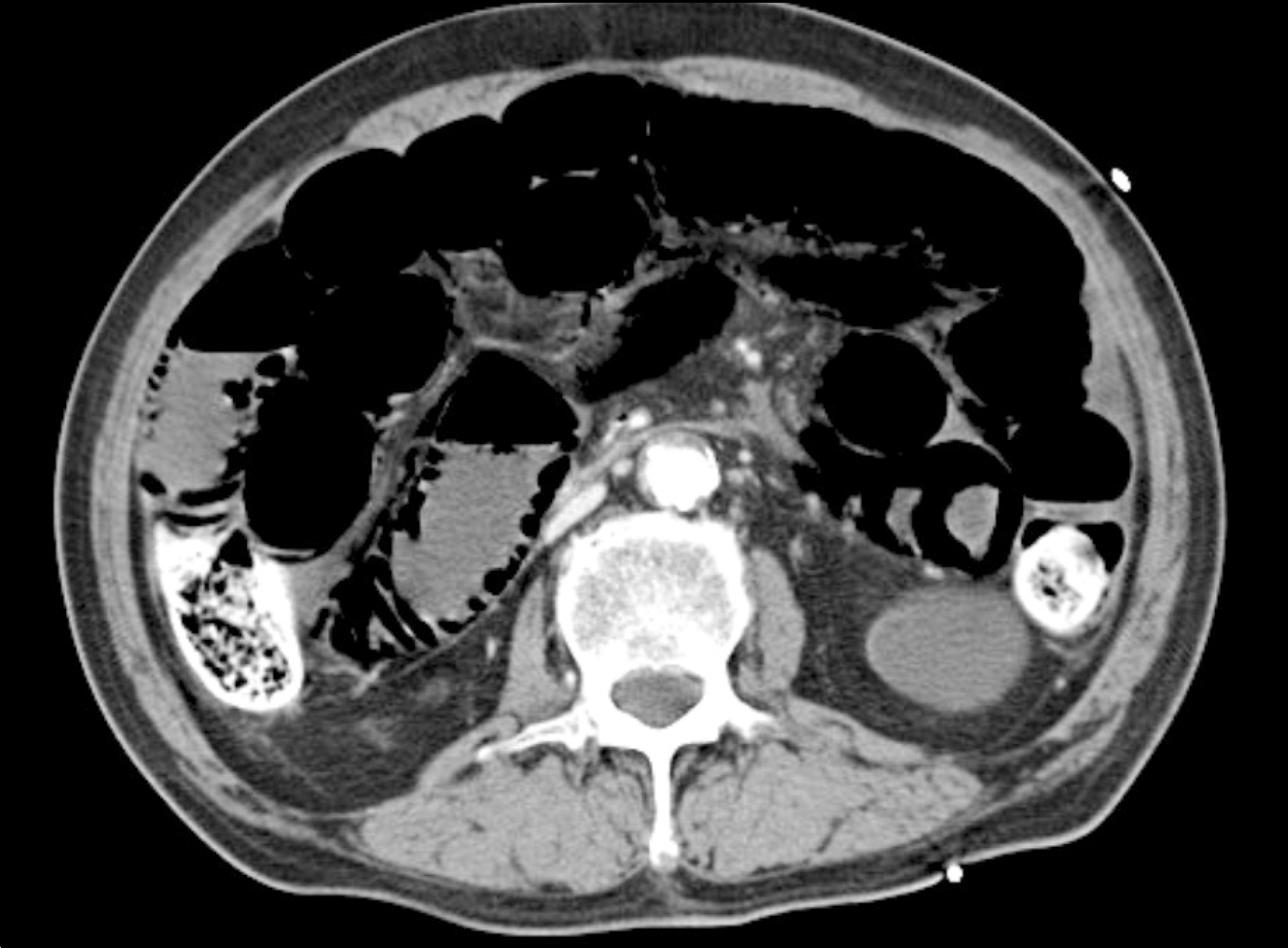
 | Fig. 3.Contrast enhanced CT scan shows intramural linear gas-like pneumatosis intestinalis in the small bowel (case 2). |
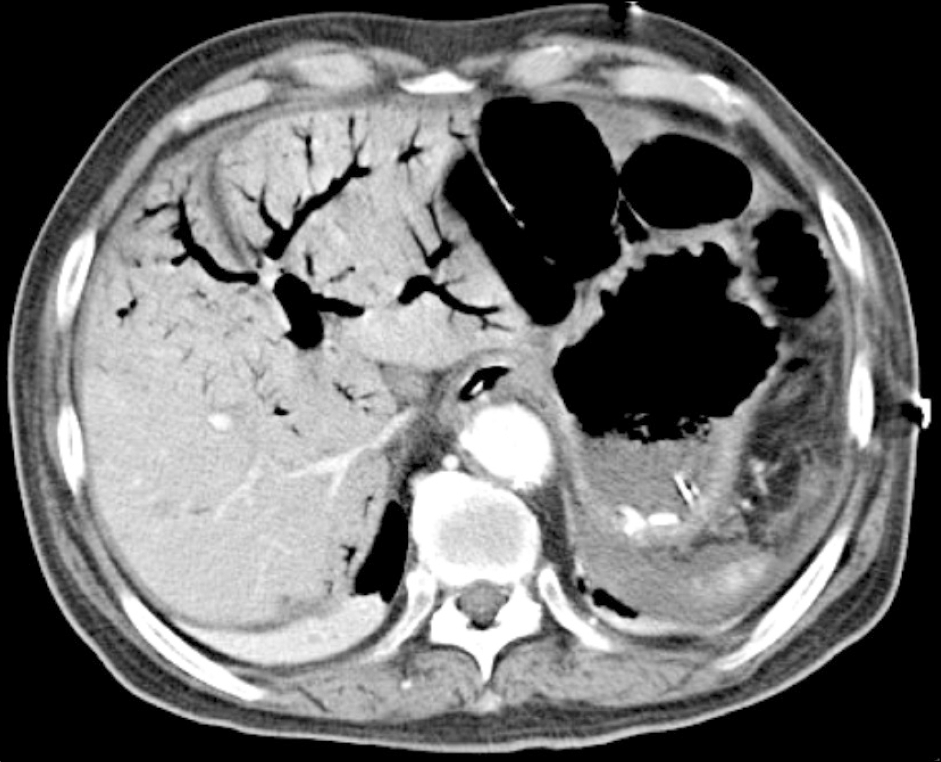
 | Fig. 4.Contrast enhanced CT scan shows large amount of gas in the intrahepatic portal vein (case 2). |
Table 1.
Clinical Characteristics of the 11 Patients with Pneumatosis Cystoides Intestinalis


 PDF
PDF ePub
ePub Citation
 Print
Print


 XML Download