Abstract
Fibrovascular polyps are rare benign intraluminal tumors that usually arise from the cervical esophagus. These often present as very large sized pedunculated polyps and cause symptoms including dysphagia and respiratory distress. Generally, large polyps are surgically excised, while endoscopic resection is limited to smaller polyps. Herein, we present a giant fibrovascular polyp of the esophagus treated successfully by endoscopic resection.
References
1. Owens JJ, Donovan DT, Alford EL, et al. Life-threatening presentations of fibrovascular esophageal and hypopharyngeal polyps. Ann Otol Rhinol Laryngol. 1994; 103:838–842.

2. Ginai AZ, Halfhide BC, Dees J, Zondervan PE, Klooswijk AI, Knegt PP. Giant esophageal polyp: a clinical and radiological entity with variable histology. Eur Radiol. 1998; 8:264–269.

3. Paik HC, Han JW, Jung EK, Bae KM, Lee YH. Fibrovascular polyp of the esophagus in infant. Yonsei Med J. 2001; 42:264–266.

4. Seshul MJ, Wiatrak BJ, Galliani CA, Odrezin GT. Pharyngeal fibrovascular polyp in a child. Ann Otol Rhinol Laryngol. 1998; 107:797–800.

5. Levine MS, Buck JL, Pantongrag-Brown L, Buetow PC, Hallman JR, Sobin LH. Fibrovascular polyps of the esophagus: clinical, radiographic, and pathologic findings in 16 patients. AJR Am J Roentgenol. 1996; 166:781–787.

6. Caceres M, Steeb G, Wilks SM, Garrett HE Jr. Large pedunculated polyps originating in the esophagus and hypopharynx. Ann Thorac Surg. 2006; 81:393–396.

7. Lee SY, Chan WH, Sivanandan R, Lim DT, Wong WK. Recurrent giant fibrovascular polyp of the esophagus. World J Gastroenterol. 2009; 15:3697–3700.

8. Ramalho LN, Martin CC, Zerbini T. Sudden death caused by fibrovascular esophageal polyp: case report and study review. Am J Forensic Med Pathol. 2010; 31:103–105.
9. Kim TS, Song SY, Han J, Shim YM, Jeong HS. Giant fibrovascular polyp of the esophagus: CT findings. Abdom Imaging. 2005; 30:653–655.

10. Ascenti G, Racchiusa S, Mazziotti S, Bottari M, Scribano E. Giant fibrovascular polyp of the esophagus: CT and MR findings. Abdom Imaging. 1999; 24:109–110.

11. Alobid I, Vilaseca I, Fernández J, Bordas JM. Giant fibrovascular polyp of the esophagus causing sudden dyspnea: endoscopic treatment. Laryngoscope. 2007; 117:944–945.

12. Chauhan S, Draganov P. Endoscopic removal of two giant fibrovascular polyps of the esophagus using the "two channel, two devices technique". Gastrointest Endosc. 2011; 73:1036–1037.

13. Di Mitri R, Mocciaro F, Lipani M, Corrao S. One-step endoscopic removal of a giant double esophageal fibrovascular polyp. Dig Liver Dis. 2014; 46:660–662.

14. Murino A, Eisendrath P, Blero D, Ibrahim M, Neuhaus H, Devière J. A giant fibrovascular esophageal polyp endoscopically resected using 2 gastroscopes simultaneously (with videos). Gastrointest Endosc. 2014; 79:834–835.

15. Park JS, Bang BW, Shin J, et al. A case of esophageal fibrovascular polyp that induced asphyxia during sleep. Clin Endosc. 2014; 47:101–103.

16. Lobo N, Hall A, Weir J, Mace A. Endoscopic resection of a giant fibrovascular polyp of the oesophagus with the assistance of ultrasonic shears. BMJ Case Rep. 2016. DOI: doi:10.1136/bcr-2015–214158.

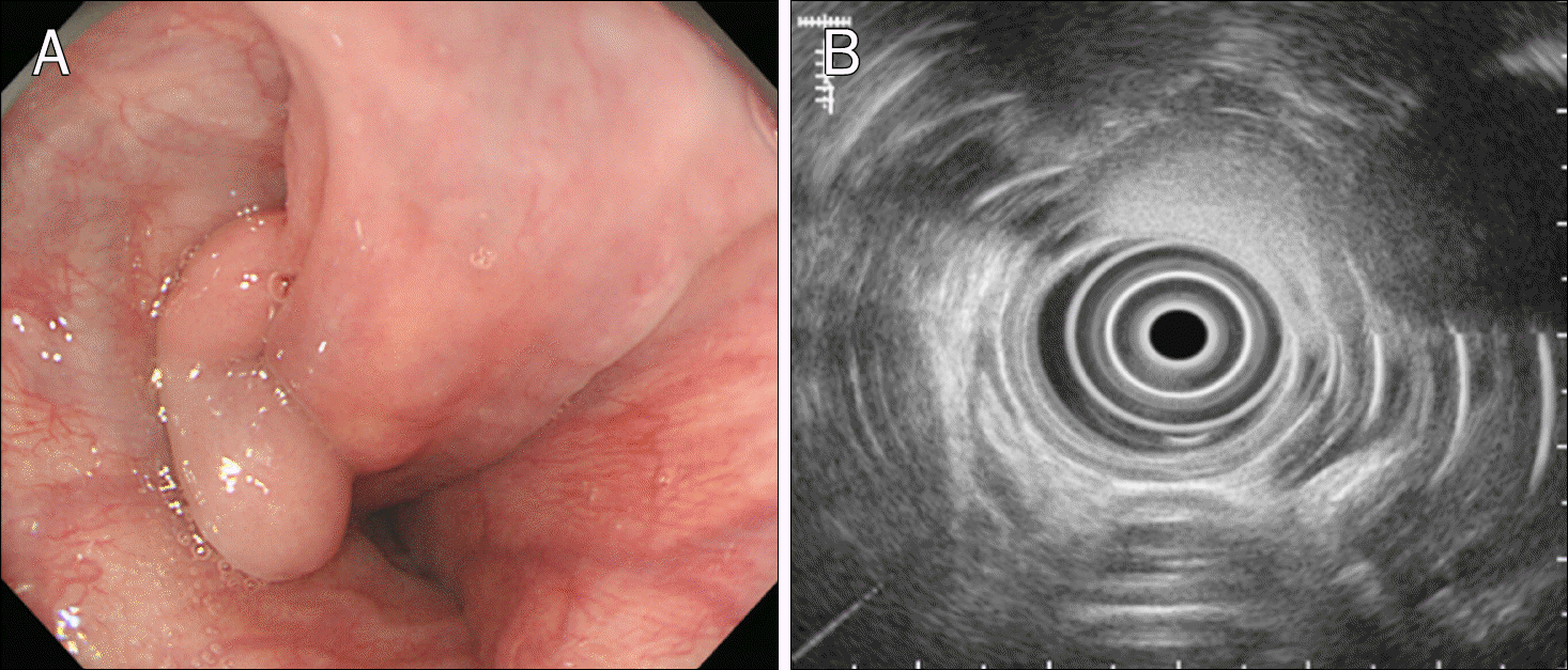
Fig. 1.
Endoscopic and endosonographic findings. (A) An about 10 cm sized sausage-shaped pedunculated polyp arises from the cervical esophagus. (B) Endosonography shows a hyperechoic homogenous lesion with clear margin on the third layer of the esophagus.

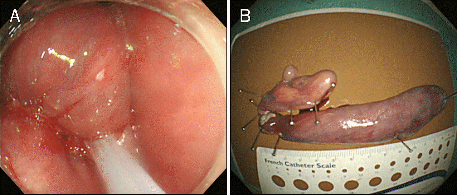
Fig. 2.
Endoscopic resection and macroscopic findings. (A) Endoscopic resection is done with a polypectomy snare. (B) The resected specimen is 12.5×3.2×1.5 cm.

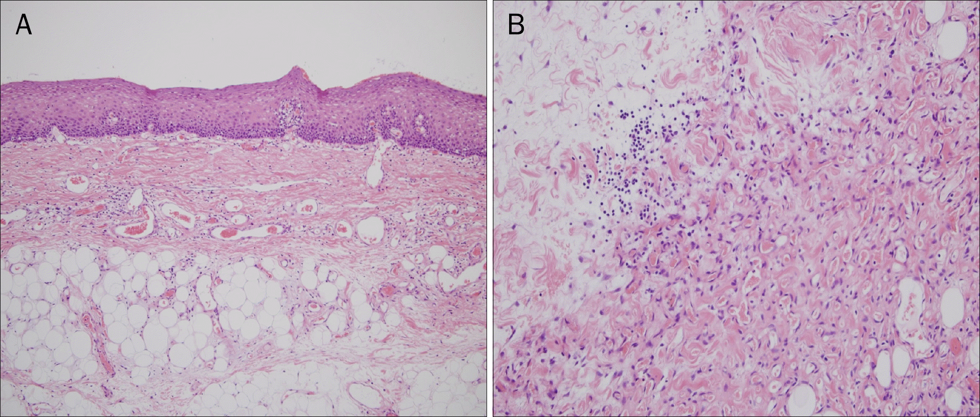
Fig. 3.
Histopathologic findings (H&E). (A) The polyp is covered with benign squamous epithelium. The core of the polyp is composed of loose fibrous tissues, adipose cells, and blood vessels (×100). (B) On high magnifying view, spindle cells set in collagenous matrix, scattered small blood vessels and inflammatory cells are seen (×400).


 PDF
PDF ePub
ePub Citation
 Print
Print


 XML Download