Abstract
Background/Aims
This study evaluated the diagnostic efficacy of fluorine-18 fluorodeoxyglucose PET/CT (F-18 FDG PET/CT) for patients with gastric mucosa-associated lymphoid tissue (MALT) lymphoma and examined the association between FDG avidity and the clinical factors affecting lesions.
Methods
Among the patients diagnosed with gastric MALT lymphoma, 16 who underwent a PET/CT for gastric MALT lymphoma were semi-quantitatively and qualitatively tested for FDG avidity of lesions in the stomach. Retrospectively collected data was analyzed to investigate the clinicoradiological factors and endoscopic findings between the patients with positive F-18 FDG PET/CT scans and those with negative scans.
Results
Eight of the 16 patients showed FDG avidity. When comparing the size of lesions in the stomach, the patients with FDG avidity had significantly larger lesions than those without (28.8 mm vs. 15.0 mm, p=0.03). The FDG-avid group has a significantly higher rate of positive CT scans than the non-avid group (75% vs. 13%, p=0.03). According to the endoscopic finding of the lesions, FDG avidity was pronounced with 75% of the protruding tumors, and 100% of the erosive-ulcerative types, which are a type of depressed tumors.
Conclusions
When gastric MALT lymphoma is large, when lesions are found using abdominal CT scans, and the macroscopic appearance of a lesion is that of a protruding tumor or erosive-ulcerative type of depressed tumor, there is a high probability that such patients may have a positive F-18 FDG PET/CT scan.
Go to : 
References
1. Radan L, Fischer D, Bar-Shalom R, et al. FDG avidity and PET/CT patterns in primary gastric lymphoma. Eur J Nucl Med Mol Imaging. 2008; 35:1424–1430.

2. Radaszkiewicz T, Dragosics B, Bauer P. Gastrointestinal malignant lymphomas of the mucosa-associated lymphoid tissue: factors relevant to prognosis. Gastroenterology. 1992; 102:1628–1638.

3. Taal BG, Burgers JM, van Heerde P, Hart AA, Somers R. The clinical spectrum and treatment of primary non-Hodgkin's lymphoma of the stomach. Ann Oncol. 1993; 4:839–846.

4. Taal BG, Boot H, van Heerde P, de Jong D, Hart AA, Burgers JM. Primary non-Hodgkin lymphoma of the stomach: endoscopic pattern and prognosis in low versus high grade malignancy in relation to the MALT concept. Gut. 1996; 39:556–561.

5. Fung CY, Grossbard ML, Linggood RM, et al. Mucosa-associated lymphoid tissue lymphoma of the stomach: long term outcome after local treatment. Cancer. 1999; 85:9–17.
6. Toyoda H, Ono T, Kiyose M, et al. Gastric mucosa-associated lymphoid tissue lymphoma with a focal high-grade component diagnosed by EUS and endoscopic mucosal resection for histologic evaluation. Gastrointest Endosc. 2000; 51:752–755.

7. Queneau PE, Helg C, Brundler MA, et al. Diagnosis of a gastric mucosa-associated lymphoid tissue lymphoma by endoscopic ultrasonography-guided biopsies in a patient with a parotid gland localization. Scand J Gastroenterol. 2002; 37:493–496.

8. Fujishima H, Misawa T, Maruoka A, Chijiiwa Y, Sakai K, Nawata H. Staging and follow-up of primary gastric lymphoma by endoscopic ultrasonography. Am J Gastroenterol. 1991; 86:719–724.
9. Lévy M, Hammel P, Lamarque D, et al. Endoscopic ultrasonography for the initial staging and follow-up in patients with low-grade gastric lymphoma of mucosa-associated lymphoid tissue treated medically. Gastrointest Endosc. 1997; 46:328–333.

10. Grau E, Gomez A, Cuñat A, Oltra C. Computed tomography in staging of primary gastric lymphoma. Lancet. 1996; 347:1261.

11. Delbeke D, Martin WH, Morgan DS, et al. 2-deoxy-2-[F-18]fluoro- D-glucose imaging with positron emission tomography for initial staging of Hodgkin's disease and lymphoma. Mol Imaging Biol. 2002; 4:105–114.
12. Kostakoglu L, Coleman M, Leonard JP, Kuji I, Zoe H, Goldsmith SJ. PET predicts prognosis after 1 cycle of chemotherapy in aggressive lymphoma and Hodgkin's disease. J Nucl Med. 2002; 43:1018–1027.
13. Spaepen K, Stroobants S, Dupont P, et al. Early restaging positron emission tomography with (18)F-fluorodeoxyglucose predicts outcome in patients with aggressive non-Hodgkin's lymphoma. Ann Oncol. 2002; 13:1356–1363.

14. Guay C, Lépine M, Verreault J, Bénard F. Prognostic value of PET using 18F-FDG in Hodgkin's disease for posttreatment evaluation. J Nucl Med. 2003; 44:1225–1231.
15. Ambrosini V, Rubello D, Castellucci P, et al. Diagnostic role of 18F-FDG PET in gastric MALT lymphoma. Nucl Med Rev Cent East Eur. 2006; 9:37–40.
16. Perry C, Herishanu Y, Metzer U, et al. Diagnostic accuracy of PET/CT in patients with extranodal marginal zone MALT lymphoma. Eur J Haematol. 2007; 79:205–209.

17. Beal KP, Yeung HW, Yahalom J. FDG-PET scanning for detection and staging of extranodal marginal zone lymphomas of the MALT type: a report of 42 cases. Ann Oncol. 2005; 16:473–480.

18. Hoffmann M, Wöhrer S, Becherer A, et al. 18F-Fluoro-de-oxy-glucose positron emission tomography in lymphoma of mu-cosa-associated lymphoid tissue: histology makes the difference. Ann Oncol. 2006; 17:1761–1765.

19. Alinari L, Castellucci P, Elstrom R, et al. 18F-FDG PET in mucosa-associated lymphoid tissue (MALT) lymphoma. Leuk Lymphoma. 2006; 47:2096–2101.

20. Enomoto K, Hamada K, Inohara H, et al. Mucosa-associated lymphoid tissue lymphoma studied with FDG-PET: a comparison with CT and endoscopic findings. Ann Nucl Med. 2008; 22:261–267.

Go to : 
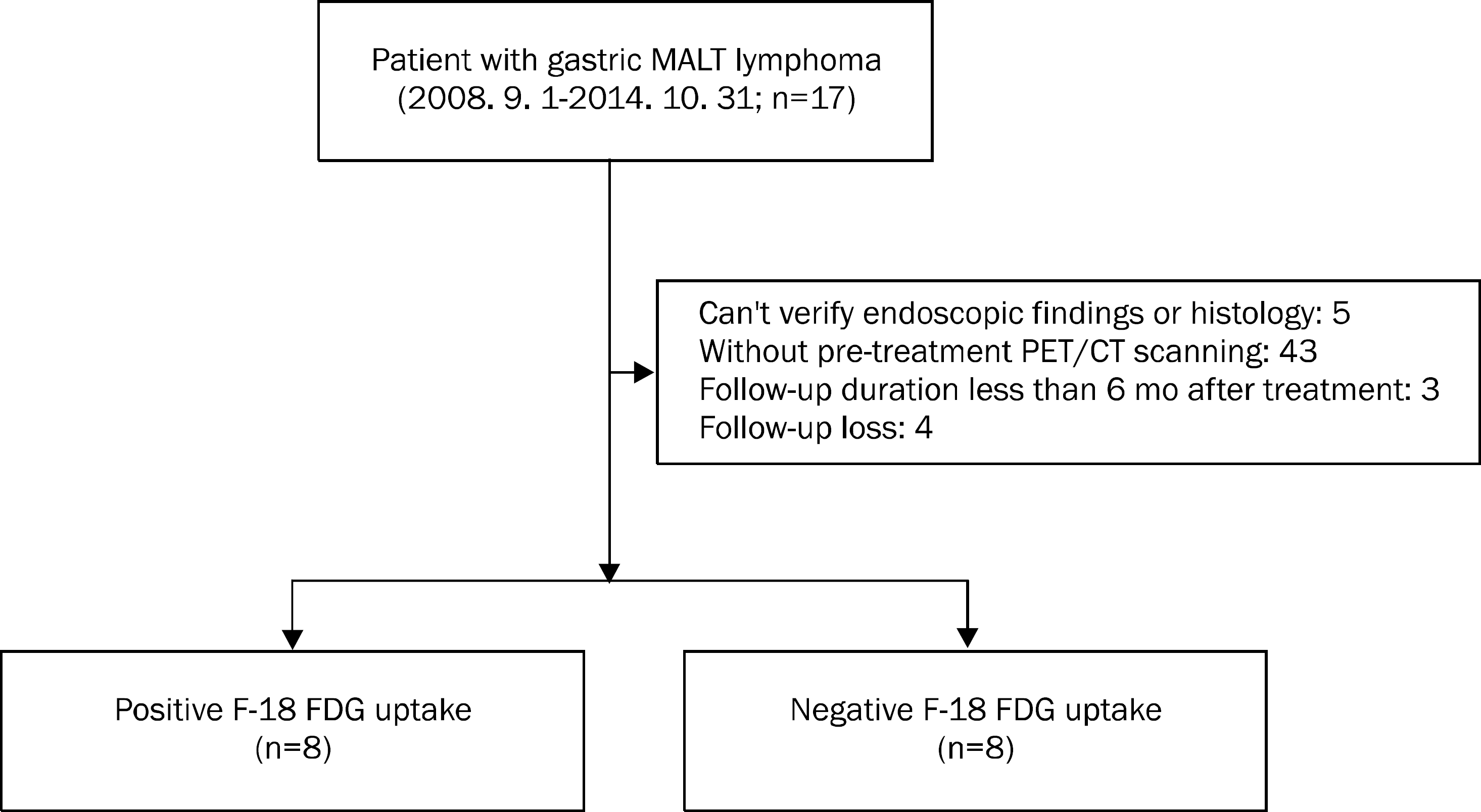
 | Fig. 1.Flowchart shows patient inclusion. MALT, mucosa-associated lymphoid tissue; F-18 FDG, fluorine-18 fluorodeoxyglucose. |
 | Fig. 2.This case shows the fluorine-18 fluorodeoxyglucose (F-18 FDG) uptake in an erosive-ulcerative type of gastric mucosa-associated lymphoid tissue lymphoma. (A) A 56-year-old female patient had an erosive-ulcerative lesion on the antral posterior wall of the stomach. (B) Maximum standardized uptake value of F-18 FDG uptake (arrow) was 6.2. (C) Abdomen CT shows irregular wall thickening at the gastric antrum. |
 | Fig. 3.This case shows no fluorine-18 fluorodeoxyglucose (F-18 FDG) uptake in a discolored-scar type of gastric mucosa-associated lymphoid tissue lymphoma. (A) A 53-year-old male patient had a discolored-scar lesion on the greater curvature in lower body of the stomach. (B) F-18 FDG PET revealed no significant F-18 FDG uptake in the stomach. (C) Abdomen CT shows no wall thickening at the gastric body. |
Table 1.
Comparison between Patients with Positive F-18 FDG Uptake and Patients with Negative F-18 FDG Uptake
Table 2.
Endoscopic Classification of Gastric MALT Lymphoma and F-18 FDG Uptake Patterns


 PDF
PDF ePub
ePub Citation
 Print
Print


 XML Download