Abstract
Pseudomyxoma peritonei is a very rare condition, and even rarer in patients with history of cancer. A 70-year old woman with a history of breast cancer was admitted with abdominal pain and distention. Abdominal computed tomography revealed ascites collection, diffuse engorgement and infiltration of the mesenteric vessel, suggesting peritonitis or peritoneal carcinomatosis. Diagnostic paracentesis was attempted several times, but a sufficient specimen could not be collected due to the thick and gelatinous nature of the ascites. Therefore, the patient underwent diagnostic laparoscopy for tissue biopsy of the peritoneum, which indicated pseudomyxoma peritonei. However, the origin of the pseudomyxoma peritonei could not be identified intraoperatively due to adhesions and large amount of mucoceles. Systemic chemotherapy was performed using Fluorouracil, producing some symptomatic relief. After discharge, abdominal pain and distention gradually worsened, so at 18 months after initial diagnosis the patient received palliative surgery based on massive mucinous ascites and palpable mass at the omentum. The patient expired after surgery due to massive bleeding.
Go to : 
References
1. Smeenk RM, van Velthuysen ML, Verwaal VJ, Zoetmulder FA. Appendiceal neoplasms and pseudomyxoma peritonei: a population based study. Eur J Surg Oncol. 2008; 34:196–201.

2. Mann WJ Jr, Wagner J, Chumas J, Chalas E. The management of pseudomyxoma peritonei. Cancer. 1990; 66:1636–1640.

3. Smeenk RM, Bruin SC, van Velthuysen ML, Verwaal VJ. Pseudomyxoma peritonei. Curr Probl Surg. 2008; 45:527–575.

5. Fann JI, Vierra M, Fisher D, Oberhelman HA Jr, Cobb L. Pseudomyxoma peritonei. Surg Gynecol Obstet. 1993; 177:441–447.
6. Dixit A, Robertson JH, Mudan SS, Akle C. Appendiceal muco-coeles and pseudomyxoma peritonei. World J Gastroenterol. 2007; 13:2381–2384.

7. Carmignani CP, Hampton R, Sugarbaker CE, Chang D, Sugarbaker PH. Utility of CEA and CA 19–9 tumor markers in diagnosis and prognostic assessment of mucinous epithelial cancers of the appendix. J Surg Oncol. 2004; 87:162–166.
8. Sugarbaker PH, Ronnett BM, Archer A, et al. Pseudomyxoma peritonei syndrome. Adv Surg. 1996; 30:233–280.
9. Zhou ML, Yan FH, Xu PJ, Zhang LJ, Li QH, Ji Y. Mucinous cystadenoma of the appendix: CT findings. Chin Med J (Engl). 2006; 119:1300–1303.

10. Wang H, Chen YQ, Wei R, et al. Appendiceal mucocele: A diagnostic dilemma in differentiating malignant from benign lesions with CT. AJR Am J Roentgenol. 2013; 201:W590–W595.

11. Sulkin TV, O'Neill H, Amin AI, Moran B. CT in pseudomyxoma peritonei: a review of 17 cases. Clin Radiol. 2002; 57:608–613.

12. Kreel L, Bydder GM. Computed tomography of fluid collections within the abdomen. J Comput Tomogr. 1980; 4:105–115.

13. Low RN, Barone RM, Gurney JM, Muller WD. Mucinous appendiceal neoplasms: preoperative MR staging and classification compared with surgical and histopathologic findings. AJR Am J Roentgenol. 2008; 190:656–665.

14. Zanati SA, Martin JA, Baker JP, Streutker CJ, Marcon NE. Colonoscopic diagnosis of mucocele of the appendix. Gastrointest Endosc. 2005; 62:452–456.

15. Caskey CI, Scatarige JC, Fishman EK. Distribution of metastases in breast carcinoma: CT evaluation of the abdomen. Clin Imaging. 1991; 15:166–171.

16. Chua TC, Moran BJ, Sugarbaker PH, et al. Early- and long-term outcome data of patients with pseudomyxoma peritonei from appendiceal origin treated by a strategy of cytoreductive surgery and hyperthermic intraperitoneal chemotherapy. J Clin Oncol. 2012; 30:2449–2456.
17. Sugarbaker PH. New standard of care for appendiceal epithelial neoplasms and pseudomyxoma peritonei syndrome? Lancet Oncol. 2006; 7:69–76.

18. Glehen O, Mohamed F, Sugarbaker PH. Incomplete cytor-eduction in 174 patients with peritoneal carcinomatosis from appendiceal malignancy. Ann Surg. 2004; 240:278–285.

Go to : 
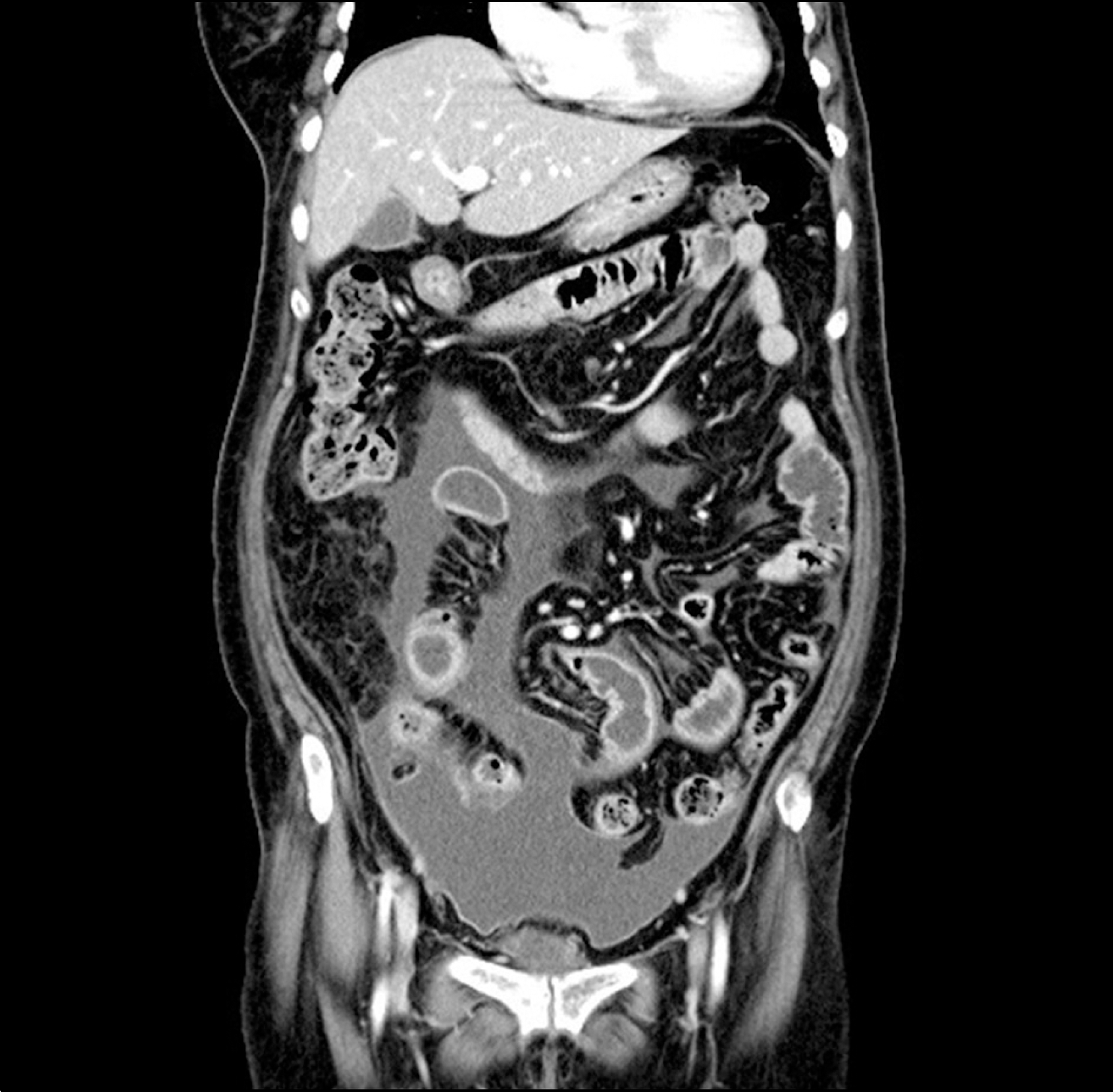
 | Fig. 1.Abdominopelvic CT findings on the day of admission. Ascites collection in the lower abdomen, pelvic cavity with diffuse peritoneal enhancement and segmental wall thickening of the distal ileal loops. |
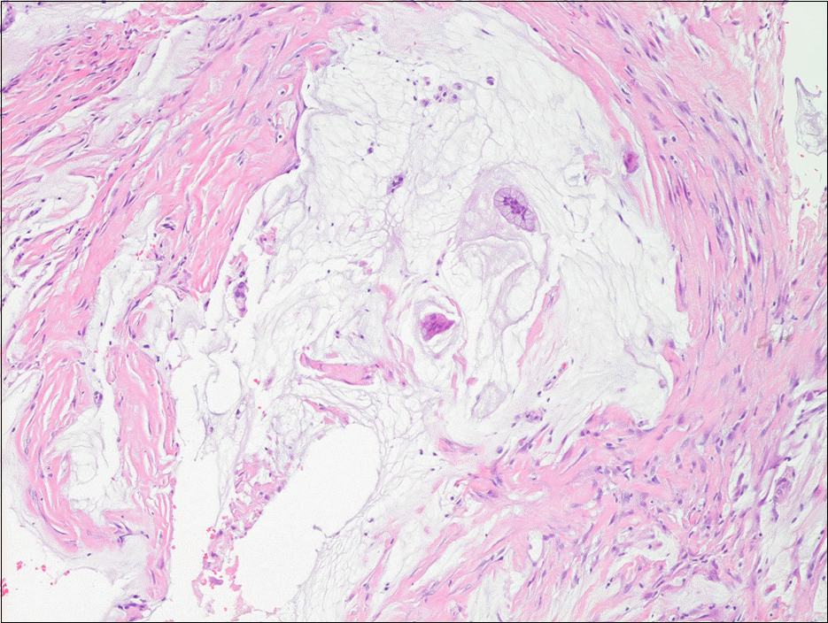
 | Fig. 2.Biopsy on peritoneal tissues taken from laparoscopy. Pools of mucin with a few clusters of mucinous columnar epithelium (H&E, ×100). |
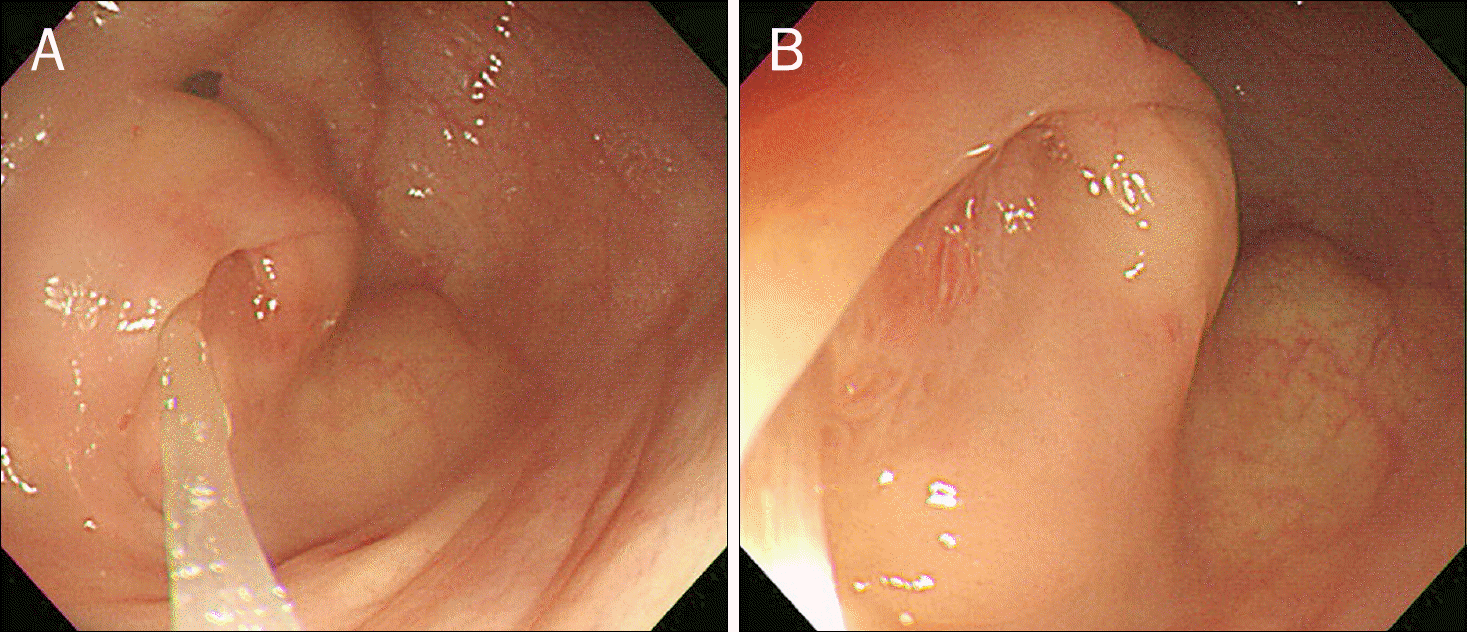
 | Fig. 3.Colonoscopic findings of the orifice of appendix. (A) Colonoscopy showing mucin leaking out from the appendix orifice and (B) mucoceles on the appendix orifice. |
 | Fig. 4.Abdominopelvic CT findings before the palliative surgery. (A) Ascites and hepatic surface scalloping and (B) soft tissue mass at the omentum. |
 | Fig. 5.Biopsy on peritoneal tissues taken from palliative surgery (H&E). (A) Peritoneal involvement of mucinous neoplasm (×40). (B) Abundant extracellular mucin with strips of low grade mucinous epithelium (×200). (C) Foci of mucinous epithelium with high grade cytologic atypia were identified. Findings are consistent with mucinous carcinoma peritonei (×200). |


 PDF
PDF ePub
ePub Citation
 Print
Print


 XML Download