Abstract
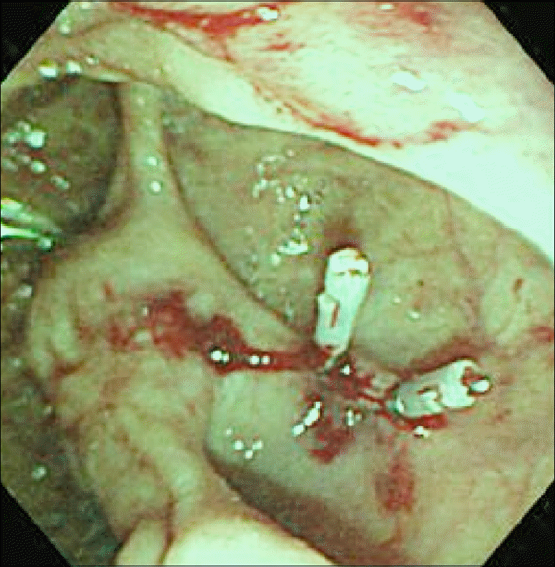
Periampullary diverticulum is commonly found during endoscopy and can occur at any age although its prevalence increases with age. Periampullary diverticular bleeding is a rare and difficult to diagnose during clinical practice because of its unique appearance and location. This often can lead to massive bleeding and interfere with adequate bleeding control. Endoscopic management on duodenal diverticular bleeding is limited compared to colonic diverticular bleeding due to lack of experience. Herein, we report a case of active bleeding from a periampullary diverticulum during bile duct stone extraction diagnosed by side-viewing endoscope and successfully controlled using hemoclips without any complications.
References
1. Chen YY, Yen HH, Soon MS. Impact of endoscopy in the management of duodenal diverticular bleeding: experience of a single medical center and a review of recent literature. Gastrointest Endosc. 2007; 66:831–835.

2. Onozato Y, Kakizaki S, Ishihara H, et al. Endoscopic management of duodenal diverticular bleeding. Gastrointest Endosc. 2007; 66:1042–1049.

3. Raju GS, Nath S, Zhao X, Jafri S, Gomez G, Luthra G. Duodenal diverticular hemostasis with hemoclip placement on the bleeding and feeder vessels: A case report. Gastrointest Endosc. 2003; 57:116–117.

4. Lee WS, Cho SB, Park SY, et al. Successful side-viewing endoscopic hemoclipping for Dieulafoy-like lesion at the brim of a periampullary diverticulum. BMC Gastroenterol. 2010; 10:24.

5. Nishiyama N, Mori H, Rafiq K, et al. Active bleeding from a periampullary duodenal diverticulum that was difficult to diagnose but successfully treated using hemostatic forceps: a case report. J Med Case Rep. 2012; 6:367.

6. Ko KH, Lee SY, Hong SP, Hwang SK, Park PW, Rim KS. Duodenal perforation after endoscopic hemoclip application for bleeding from Dieulafoy's lesion in a duodenal diverticulum. Gastrointest Endosc. 2005; 62:781–782. discussion 782.

7. Yin WY, Chen HT, Huang SM, Lin HH, Chang TM. Clinical analysis and literature review of massive duodenal diverticular bleeding. World J Surg. 2001; 25:848–855.

8. Afridi SA, Fichtenbaum CJ, Taubin H. Review of duodenal diverticula. Am J Gastroenterol. 1991; 86:935–938.
9. Seneviratne SA, Samarasekera DN. Massive gastrointestinal haemorrhage from a duodenal diverticulum: a case report. Cases J. 2009; 2:6710.

10. Rajnakova A, Goh PM, Ngoi SS, Lim SG. ERCP in patients with periampullary diverticulum. Hepatogastroenterology. 2003; 50:625–628.
11. Khandelwal M, Akerman PA, Jones WF, Haber GB. Endoscopic therapy of a bleeding duodenal diverticulum. Am J Gastroenterol. 1995; 90:1328–1329.
12. Sim EK, Goh PM, Isaac JR, Kang JY, Gangaraju CR, Ti TK. Endoscopic management of a bleeding duodenal diverticulum. Gastrointest Endosc. 1991; 37:634.
13. Choudari CP, Luman W, Eastwood MA, Palmer KR. Bleeding duodenal diverticulum. Endoscopy. 1995; 27:284.

14. Dalal AA, Rogers SJ, Cello JP. Endoscopic management of hemorrhage from a duodenal diverticulum. Gastrointest Endosc. 1998; 48:418–420.

15. Lin LF, Siauw CP, Ho KS, Tung JN. Hemoclip treatment for post-endoscopic sphincterotomy bleeding. J Chin Med Assoc. 2004; 67:496–499.
Fig. 1.
Abdominal computed tomography reveals gall bladder stones (A) and proximal extrahepatic bile duct dilatation with distal common bile duct stones (B). The diameter of the distal common bile duct measures about 1 cm.
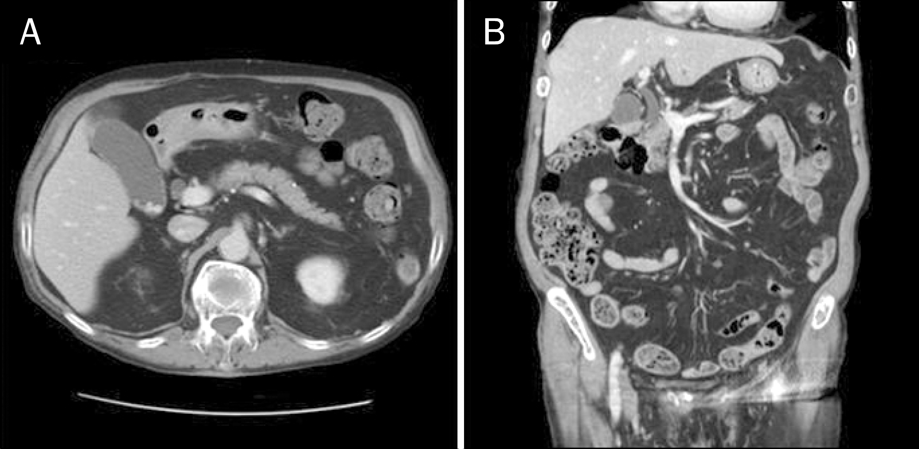
Fig. 2.
Initial duodenoscopic examination reveals a large clot and fresh blood within the periampullary diverticulum on the second portion of duodenum.
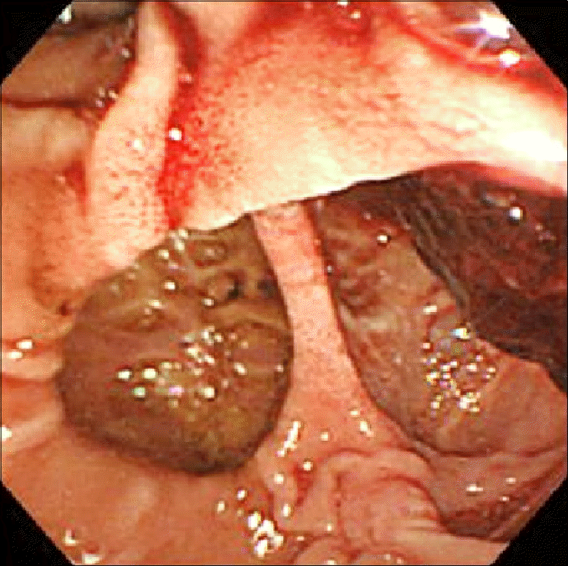
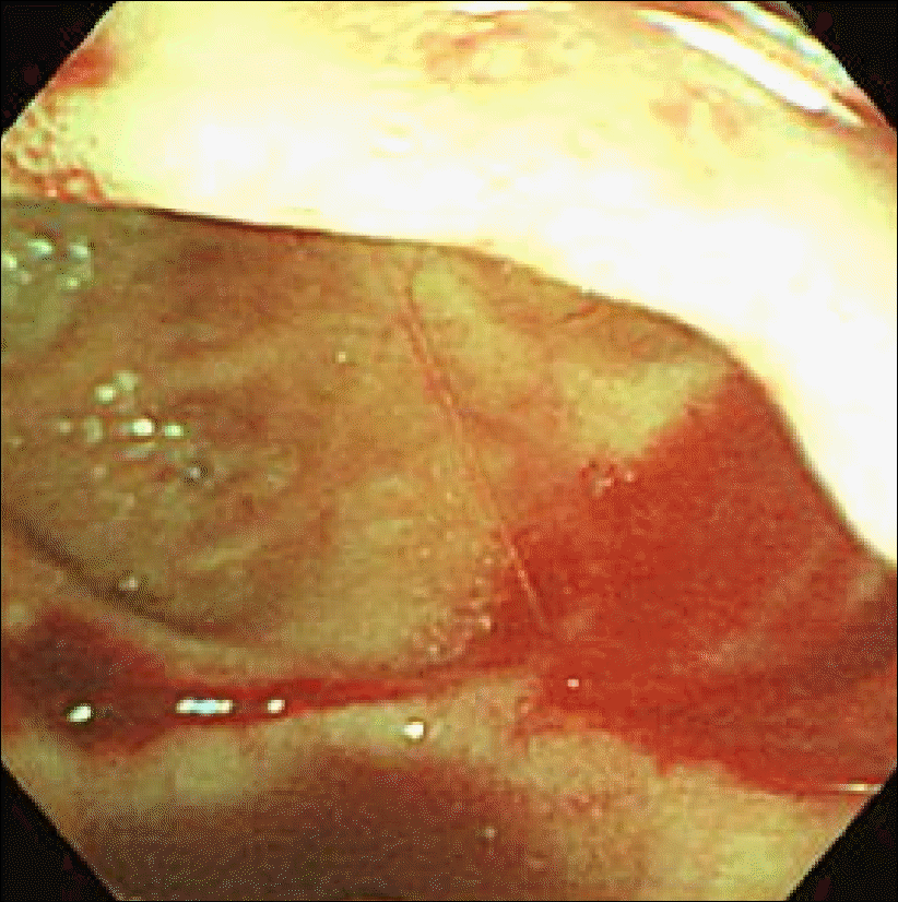
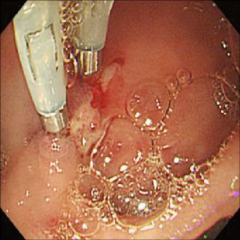
Fig. 3.
After removal of bile duct stone, spurting of blood, presumably a Dieulafoy-like lesion, was noted at the base of periampullary diverticulum.


 PDF
PDF ePub
ePub Citation
 Print
Print


 XML Download