

 PDF
PDF ePub
ePub Citation
 Print
Print


Abstract
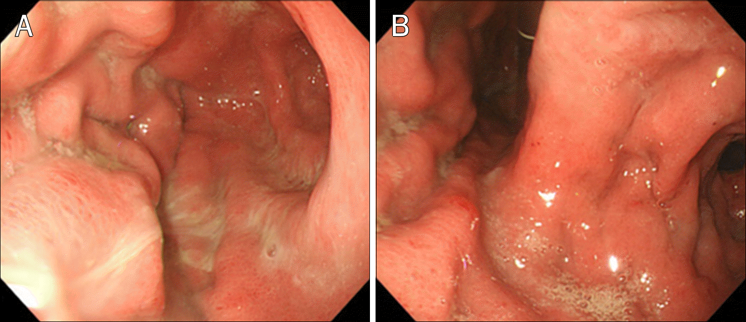
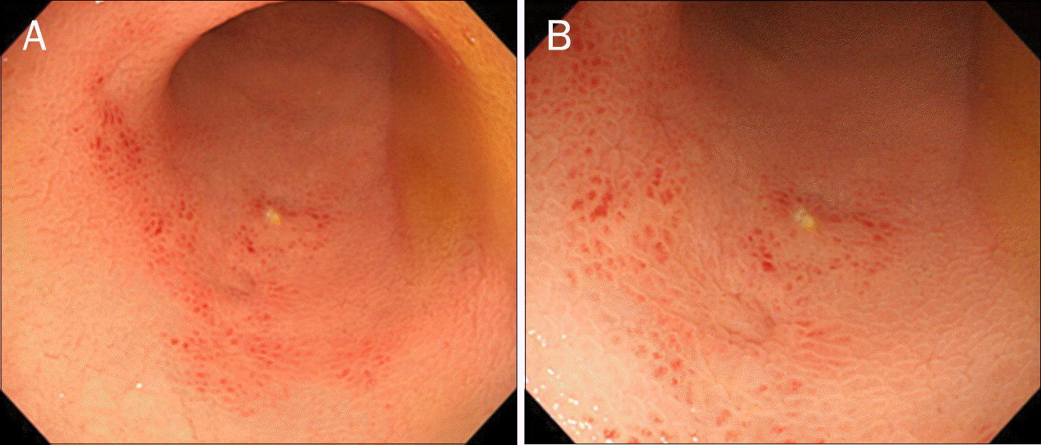
Lupus enteritis is a rare, severe complication of systemic lupus erythematosus (SLE), needing prompt diagnosis and proper management. However, SLE rarely presents as lupus enteritis at the time of initial diagnosis. Thus, delayed diagnosis and misdiagnosis are common. We report a case of a 25-year-old woman with lupus panenteritis. The patient had multiple hospitalizations for abdominal pain, nausea, and diarrhea, initially without any other symptoms suggestive of SLE, but was later observed to have malar rash and oral ulcers. Laboratory investigations were compatible with SLE, including positive antinuclear antibody (1:320) with speckled pattern. CT revealed diffuse hypodense submucosal thickening of the stomach, the entire small bowel, colon, appendix, and rectum. Treatment with high-dose corticosteroids followed by maintenance therapy with mycophenolate mofetil, hydroxychloroquine, and azathioprine resulted in clinical improvement. Diagnosis of lupus enteritis requires a high index of suspicion given the low incidence and nonspecific clinical findings.
References
1. Tian XP, Zhang X. Gastrointestinal involvement in systemic lupus erythematosus: insight into pathogenesis, diagnosis and treatment. World J Gastroenterol. 2010; 16:2971–2977.

2. Lee CK, Ahn MS, Lee EY, et al. Acute abdominal pain in systemic lupus erythematosus: focus on lupus enteritis (gastrointestinal vasculitis). Ann Rheum Dis. 2002; 61:547–550.
3. Janssens P, Arnaud L, Galicier L, et al. Lupus enteritis: from clinical findings to therapeutic management. Orphanet J Rare Dis. 2013; 8:67.
4. Sultan SM, Ioannou Y, Isenberg DA. A review of gastrointestinal manifestations of systemic lupus erythematosus. Rheumatology (Oxford). 1999; 38:917–932.
5. Smith LW, Petri M. Lupus enteritis: an uncommon manifestation of systemic lupus erythematosus. J Clin Rheumatol. 2013; 19:84–86.
6. Sran S, Sran M, Patel N, Anand P. Lupus enteritis as an initial presentation of systemic lupus erythematosus. Case Rep Gastrointest Med. 2014. DOI: doi: 10.1155/2014/962735.
7. Hoffman BI, Katz WA. The gastrointestinal manifestations of systemic lupus erythematosus: a review of the literature. Semin Arthritis Rheum. 1980; 9:237–247.
8. Hallegua DS, Wallace DJ. Gastrointestinal manifestations of systemic lupus erythematosus. Curr Opin Rheumatol. 2000; 12:379–385.
9. Chng HH, Tan BE, Teh CL, Lian TY. Major gastrointestinal manifestations in lupus patients in Asia: lupus enteritis, intestinal pseudoobstruction, and protein-losing gastroenteropathy. Lupus. 2010; 19:1404–1413.
10. Chan MR, Vasudev M, Piering WF, Ryan LM. Acute abdomen in a patient with systemic lupus erythematosus. Hospital Physician. 2005; 41:27–31.
11. Pyrpasopoulou A, Chatzimichailidou S, Aslanidis S. Vascular disease in systemic lupus erythematosus. Autoimmune Dis. 2012; 2012:876456.
12. Reissman P, Weiss EG, Teoh TA, Lucas FV, Wexner SD. Gangrenous ischemic colitis of the rectum: a rare complication of systemic lupus erythematosus. Am J Gastroenterol. 1994; 89:2234–2236.
13. Kim DH, Na HJ, Choi YR, et al. A case of extensive involvement of lupus enteritis, from small bowel to rectum. J Korean Rheum Assoc. 2007; 14:274–278.
14. Shirai T, Hirabayashi Y, Watanabe R, et al. The use of tacrolimus for recurrent lupus enteritis: a case report. J Med Case Rep. 2010; 4:150.
15. Oh JS, Kim YG, Lee SG, Lee CK, Yoo B. Successful treatment of recurrent lupus enteritis with rituximab. Lupus. 2010; 19:220–222.
Fig. 1.
CT of the patient shows moderate amount of ascites and diffuse hypodense submucosal thickening involving the stomach lower body (A), entire small bowel (B), colon (C), appendix and rectum (D). Sub-mucosal wall thickening of stomach lower body (A, arrow). Focal enhancement of right ureter (C, arrow), suspicious systemic lupus erythematosus involvement.

 XML Download