Abstract
Background/Aims
The optimal management of bleeding peptic ulcer with adherent clot remains controversial. The purpose of this study was to compare clinical outcome between endoscopic therapy and medical therapy. We also evaluated the risk factors of rebleeding in Forrest type IIB peptic ulcer.
Methods
Upper gastrointestinal (UGI) bleeding registry data from 8 hospitals in Korea between February 2011 and December 2013 were reviewed and categorized according to the Forrest classification. Patients with acute UGI bleeding from peptic ulcer with adherent clots were enrolled.
Results
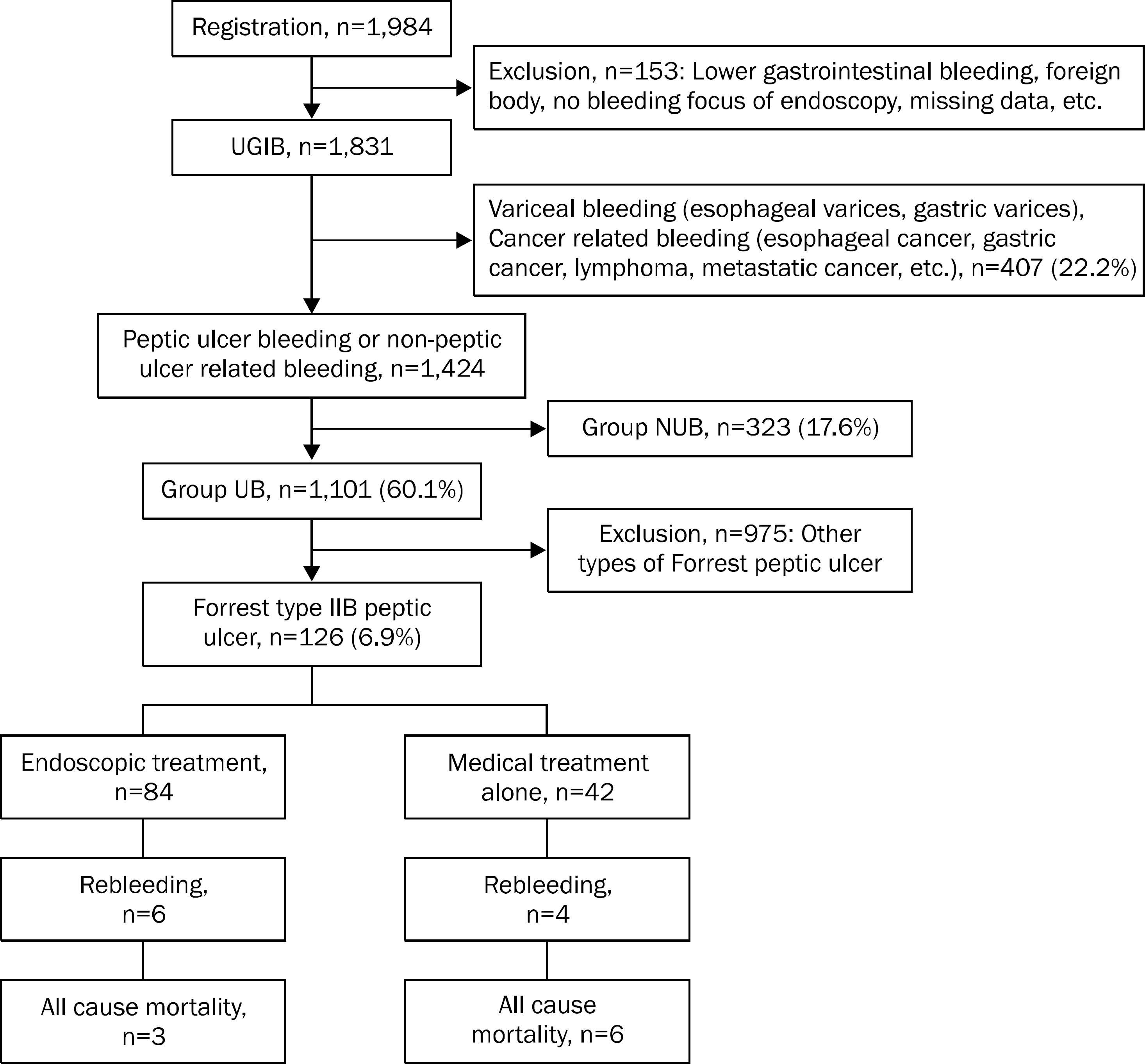
Among a total of 1,101 patients diagnosed with peptic ulcer bleeding, 126 bleedings (11.4%) were classified as Forrest type IIB. Of the 126 patients with adherent clots, 84 (66.7%) received endoscopic therapy and 42 (33.3%) were managed with medical therapy alone. The baseline characteristics of patients in two groups were similar except for higher Glasgow Blatchford Score and pre-endoscopic Rockall score in medical therapy group. Bleeding related mortality (1.2% vs.10%; p=0.018) and all cause mortality (3.7% vs. 20.0%; p=0.005) were significantly lower in the endoscopic therapy group. However, there was no difference between endoscopic therapy and medical therapy regarding rebleeding (7.1% vs. 9.5%; p=0.641). In multivariate analysis, independent risk factors of rebleeding were previous medication with aspirin and/or NSAID (OR, 13.1; p=0.025).
References
1. van Leerdam ME, Vreeburg EM, Rauws EA, et al. Acute upper GI bleeding: did anything change? Time trend analysis of incidence and outcome of acute upper GI bleeding between 1993/1994 and 2000. Am J Gastroenterol. 2003; 98:1494–1499.
2. Hearnshaw SA, Logan RF, Lowe D, Travis SP, Murphy MF, Palmer KR. Acute upper gastrointestinal bleeding in the UK: patient characteristics, diagnoses and outcomes in the 2007 UK audit. Gut. 2011; 60:1327–1335.

3. Sung JJ, Kuipers EJ, El-Serag HB. Systematic review: the global incidence and prevalence of peptic ulcer disease. Aliment Pharmacol Ther. 2009; 29:938–946.

4. Kim JI, Kim SG, Kim N, et al. Korean College of Helicobacter and Upper Gastrointestinal Research. Changing prevalence of upper gastrointestinal disease in 28 893 Koreans from 1995 to 2005. Eur J Gastroenterol Hepatol. 2009; 21:787–793.
5. Theocharis GJ, Thomopoulos KC, Sakellaropoulos G, Katsakoulis E, Nikolopoulou V. Changing trends in the epidemiology and clinical outcome of acute upper gastrointestinal bleeding in a defined geographical area in Greece. J Clin Gastroenterol. 2008; 42:128–133.

6. Hwang JH, Fisher DA, Ben-Menachem T, et al. Standards of Practice Committee of the American Society for Gastrointestinal Endoscopy. The role of endoscopy in the management of acute non-variceal upper GI bleeding. Gastrointest Endosc. 2012; 75:1132–1138.

7. Enestvedt BK, Gralnek IM, Mattek N, Lieberman DA, Eisen G. An evaluation of endoscopic indications and findings related to non-variceal upper-GI hemorrhage in a large multicenter consortium. Gastrointest Endosc. 2008; 67:422–429.
8. Proceedings of the Consensus Conference on Therapeutic Endoscopy in Bleeding Ulcers. March 6–8, 1989. Gastrointest Endosc. 1990; 36(5 Suppl):S1–S65.
10. Laine L, Stein C, Sharma V. A prospective outcome study of patients with clot in an ulcer and the effect of irrigation. Gastrointest Endosc. 1996; 43:107–110.

11. Jensen DM, Kovacs TO, Jutabha R, et al. Randomized trial of medical or endoscopic therapy to prevent recurrent ulcer hemorrhage in patients with adherent clots. Gastroenterology. 2002; 123:407–413.

12. Bleau BL, Gostout CJ, Sherman KE, et al. Recurrent bleeding from peptic ulcer associated with adherent clot: a randomized study comparing endoscopic treatment with medical therapy. Gastrointest Endosc. 2002; 56:1–6.

13. Kahi CJ, Jensen DM, Sung JJ, et al. Endoscopic therapy versus medical therapy for bleeding peptic ulcer with adherent clot: a metaanalysis. Gastroenterology. 2005; 129:855–862.

14. Laine L. Systematic review of endoscopic therapy for ulcers with clots: can a metaanalysis be misleading? Gastroenterology. 2005; 129:2127. author reply 2127–8.

15. Laine L, McQuaid KR. Endoscopic therapy for bleeding ulcers: an evidence-based approach based on meta-analyses of randomized controlled trials. Clin Gastroenterol Hepatol. 2009; 7:33–47.

16. Jung HK, Son HY, Jung SA, et al. Comparison of oral omeprazole and endoscopic ethanol injection therapy for prevention of recurrent bleeding from peptic ulcers with nonbleeding visible vessels or fresh adherent clots. Am J Gastroenterol. 2002; 97:1736–1740.

17. Blatchford O, Murray WR, Blatchford M. A risk score to predict need for treatment for upper-gastrointestinal haemorrhage. Lancet. 2000; 356:1318–1321.

18. Vreeburg EM, Terwee CB, Snel P, et al. Validation of the Rockall risk scoring system in upper gastrointestinal bleeding. Gut. 1999; 44:331–335.

19. Park JK, Jung YD, Seo YJ, et al. Risk factors for early rebleeding after initial endoscopic hemostasis in patients with bleeding peptic ulcers. Korean J Gastrointest Endosc. 2000; 21:898–908.
20. Silverstein FE, Gilbert DA, Tedesco FJ, Buenger NK, Persing J. The national ASGE survey on upper gastrointestinal bleeding. II. Clinical prognostic factors. Gastrointest Endosc. 1981; 27:80–93.
21. Rockall TA, Logan RF, Devlin HB, Northfield TC. Risk assessment after acute upper gastrointestinal haemorrhage. Gut. 1996; 38:316–321.

22. Ohmann C, Imhof M, Ruppert C, et al. Time-trends in the epidemiology of peptic ulcer bleeding. Scand J Gastroenterol. 2005; 40:914–920.

23. Gabriel SE, Jaakkimainen L, Bombardier C. Risk for serious gastrointestinal complications related to use of nonsteroidal anti-inflammatory drugs. A metaanalysis. Ann Intern Med. 1991; 115:787–796.
Fig. 1.
Study flow showing the causes of upper gastrointestinal bleeding. The numbers in parentheses are the proportions of each group relating to patients with upper gastrointestinal bleeding (UGIB). GI, gastrointestinal; NUB, non-peptic ulcer related bleeding; UB, peptic ulcer bleeding.

Table 1.
Baseline Characteristics of 126 Patients
| Variable | Endoscopic therapy (n=84) | Medical therapy (n=42) | p-value |
|---|---|---|---|
| Age (yr) | 64±15 | 68±15 | NS |
| Sex (male: female) | 63:21 | 29:13 | NS |
| Previous peptic ulcer history | NS | ||
| None | 75 (89.3) | 36 (85.7) | |
| Gastric ulcer | 9 (10.7) | 4 (9.5) | |
| Duodenal ulcer | 0 (0) | 2 (4.8) | |
| Previous aspirin and/or NSAID history | NS | ||
| None | 58 (69.1) | 31 (73.8) | |
| Yes | 26 (31.0) | 11 (26.2) | |
| Previous warfarin and/or LMWH history | NS | ||
| None | 80 (95.2) | 42 (100) | |
| Yes | 4 (4.8) | 0 (0) | |
| Recent PPI medication history | NS | ||
| None | 77 (91.7) | 35 (83.3) | |
| Yes | 7 (8.3) | 7 (16.7) | |
| Hemoglobin <8 g/dL | NS | ||
| None | 49 (58.3) | 24 (57.1) | |
| Yes | 35 (41.7) | 18 (42.9) | |
| Platelet <150,000/mm3 | NS | ||
| None | 72 (85.7) | 37 (88.1) | |
| Yes | 12 (14.3) | 5 (11.9) | |
| Prothrombin time (sec) | NS | ||
| ≤15 | 73 (86.9) | 35 (83.3) | |
| >15 | 11 (13.1) | 7 (16.8) | |
| Systolic blood pressure <90 mmHg | NS | ||
| None | 11 (13.1) | 5 (11.9) | |
| Yes | 73 (86.9) | 37 (88.1) | |
| Heart rate ≥110 beats/min | |||
| None | 72 (85.7) | 37 (88.1) | NS |
| Yes | 12 (14.3) | 5 (11.9) | |
| Endoscopic findings | NS | ||
| Gastric ulcer | 66 (78.6) | 33 (78.6) | |
| Duodenal ulcer | 18 (21.4) | 9 (21.4) | |
| Helicobacter pylori infection | NS | ||
| None | 37 (61.7) | 13 (54.17) | |
| Yes | 23 (38.3) | 11 (45.83) | |
| Glasgow Blatchford score a | 12 (1–20) | 13 (0–17) | 0.004 |
| Pre-endoscopic Rockall score b | 1 (0–7) | 3 (0–10) | 0.006 |
| Full Rockall score c | 4.5 (0–10) | 6 (1–10) | NS |
a Calculated with admission risk marker: blood urea nitrogen (0, 2, 3, 4, 6 points), hemoglobin value (0, 1, 3, 6 points for men and 0, 1, 6 points for women), systolic blood pressure (0 to 3 points) and other markers such as pulse rate (0 to 1 point), presentation with melena (1 point), presentation with syncope (2 points), hepatic disease (2 points), cardiac failure (2 points).
Table 2.
Comparison on Outcomes between Endoscopic Therapy and Medical Therapy
| Variable | Endoscopic therapy (n=84) | Medical therapy (n=42) | p-value |
|---|---|---|---|
| Rebleeding | 6 (7.1) | 4 (9.5) | NS |
| Aggravation of comorbidity a | 3 (3.6) | 5 (11.9) | NS |
| Mortality | |||
| Bleeding related mortality | 1 (1.2) | 3 (7.1) | 0.018 |
| Non bleeding related mortality | 2 (2.4) | 3 (7.1) | |
| All cause mortality | 3 (3.6) | 6 (14.3) | 0.005 |
Table 3.
Univariate Analysis of Risk Factors for Rebleeding
Table 4.
Multivariate Analysis of Risk Factors for Rebleeding
| Variable | OR (95% CI for OR) | p-value |
|---|---|---|
| Previous aspirin and/or NSAID history | ||
| None | 1 | − |
| Yes | 13.1 (1.4–124.2) | 0.025 |
| Pre-endoscopic Rockall score | 2.3 (0.7–7.5) | NS |
| Full Rockall score | 1.6 (0.4–5.6) | NS |


 PDF
PDF ePub
ePub Citation
 Print
Print


 XML Download