Abstract
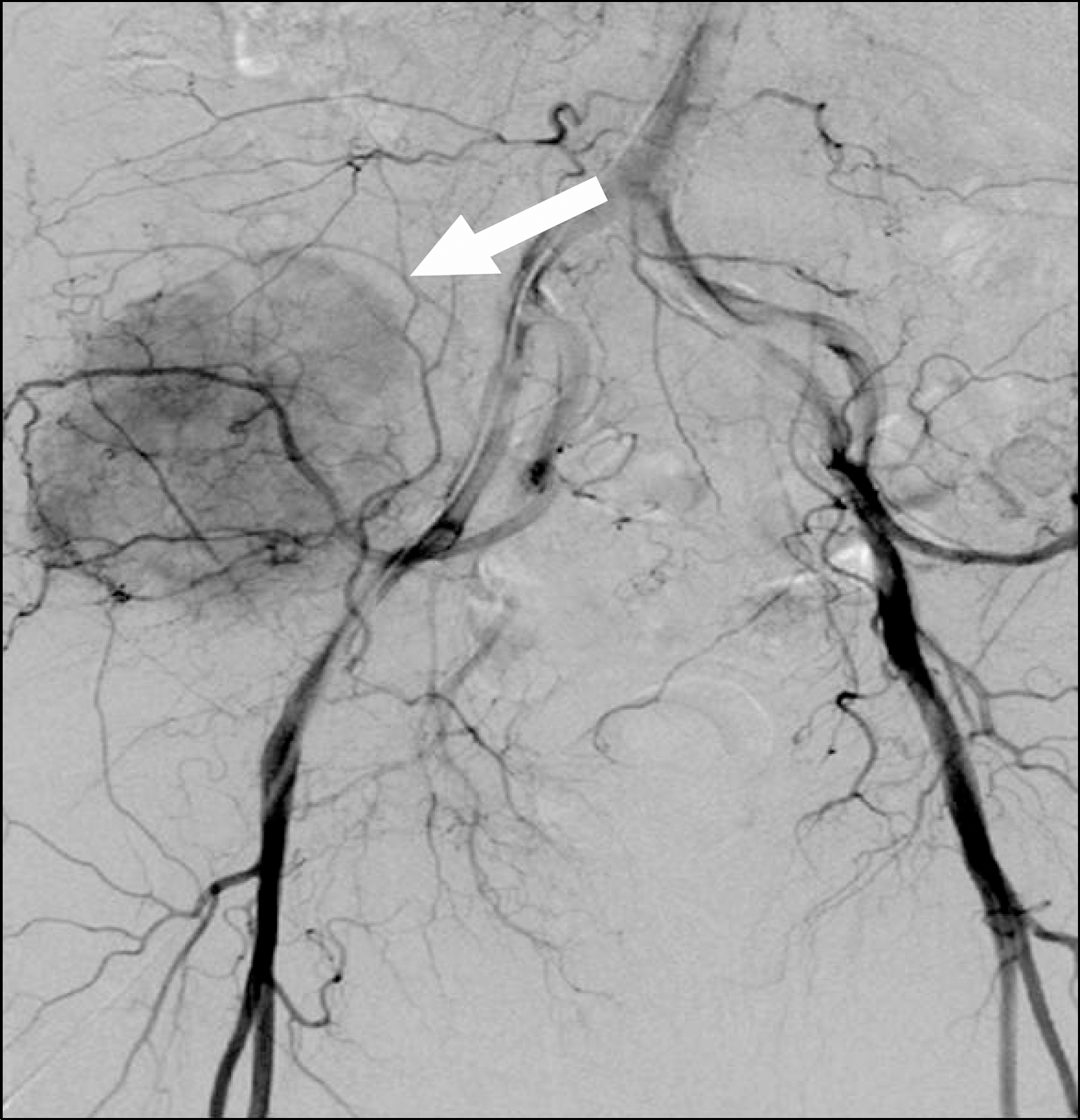
The occurrence of hepatocellular carcinoma (HCC) is closely associated with viral hepatitis or alcoholic hepatitis. Although active surveillance is ongoing in Korea, advanced or metastatic HCC is found at initial presentation in many patients. Metastatic HCC presents with a hypervascular intrahepatic tumor and extrahepatic lesions such as lung or lymph node metastases. Cases of HCC presenting as carcinoma of unknown primary have been rarely reported. The authors experienced a case of metastatic HCC in a patient who presented with a metastatic bone lesion but no primary intrahepatic tumor. This case suggests that HCC should be considered as a differential diagnosis when evaluating the primary origin of metastatic carcinoma.
References
1. Jung KW, Won YJ, Kong HJ, Oh CM, Seo HG, Lee JS. Prediction of cancer incidence and mortality in Korea, 2013. Cancer Res Treat. 2013; 45:15–21.

2. de Lope CR, Tremosini S, Forner A, Reig M, Bruix J. Management of HCC. J Hepatol. 2012; 56(Suppl 1):S75–S87.

3. Kummar S, Shafi NQ. Metastatic hepatocellular carcinoma. Clin Oncol (R Coll Radiol). 2003; 15:288–294.

4. Fukutomi M, Yokota M, Chuman H, et al. Increased incidence of bone metastases in hepatocellular carcinoma. Eur J Gastroenterol Hepatol. 2001; 13:1083–1088.

5. Natsuizaka M, Omura T, Akaike T, et al. Clinical features of hepatocellular carcinoma with extrahepatic metastases. J Gastroenterol Hepatol. 2005; 20:1781–1787.

6. Ahmad Z, Nisa AU, Uddin Z, Azad NS. Unusual metastases of hepatocellular carcinoma (hcc) to bone and soft tissues of lower limb. J Coll Physicians Surg Pak. 2007; 17:222–223.
7. Takahama J, Taoka T, Marugami N, et al. Hepatocellular carcinoma of the iliac bone with unknown primary. Skeletal Radiol. 2010; 39:721–724.

8. Jung KS, Park KH, Chon YE, et al. A case of isolated metastatic hepatocellular carcinoma arising from the pelvic bone. Korean J Hepatol. 2012; 18:89–93.

9. Hofmann HS, Spillner J, Hammer A, Diez C. A solitary chest wall metastasis from unknown primary hepatocellular carcinoma. Eur J Gastroenterol Hepatol. 2003; 15:557–559.

10. Hyun YS, Choi HS, Bae JH, et al. Chest wall metastasis from unknown primary site of hepatocellular carcinoma. World J Gastroenterol. 2006; 12:2139–2142.

11. Ahmad K, Sah P, Dhungel K, et al. An unusual case report of hepatocellular carcinoma metastasis to the chest wall presenting as breast lump. Nepal J Medi Sci. 2012; 1:138–140.

12. Oquiñena S, Guillen-Grima F, Iñarrairaegui M, Zozaya JM, Sangro B. Spontaneous regression of hepatocellular carcinoma: a systematic review. Eur J Gastroenterol Hepatol. 2009; 21:254–257.

13. Huz JI, Melis M, Sarpel U. Spontaneous regression of hepatocellular carcinoma is most often associated with tumour hypoxia or a systemic inflammatory response. HPB (Oxford). 2012; 14:500–505.

14. Sasaki T, Fukumori D, Yamamoto K, Yamamoto F, Igimi H, Yamashita Y. Management considerations for purported spontaneous regression of hepatocellular carcinoma: a case report. Case Rep Gastroenterol. 2013; 7:147–152.

15. Terracciano LM, Glatz K, Mhawech P, et al. Hepatoid adenocarcinoma with liver metastasis mimicking hepatocellular carcinoma: an immunohistochemical and molecular study of eight cases. Am J Surg Pathol. 2003; 27:1302–1312.
Fig. 3.
Biopsy specimen of the iliac bone. H&E stain shows round, eosinophilic cytoplasm-rich cells with a trabecular arrangement, suggestive of hepatocellular carcinoma (A, ×100; B, ×400). The tumor cells showed focally positive immunoreactivity for hepatocyte surface antigen (C) and CD10 (D) with a canalicular staining pattern (arrows).
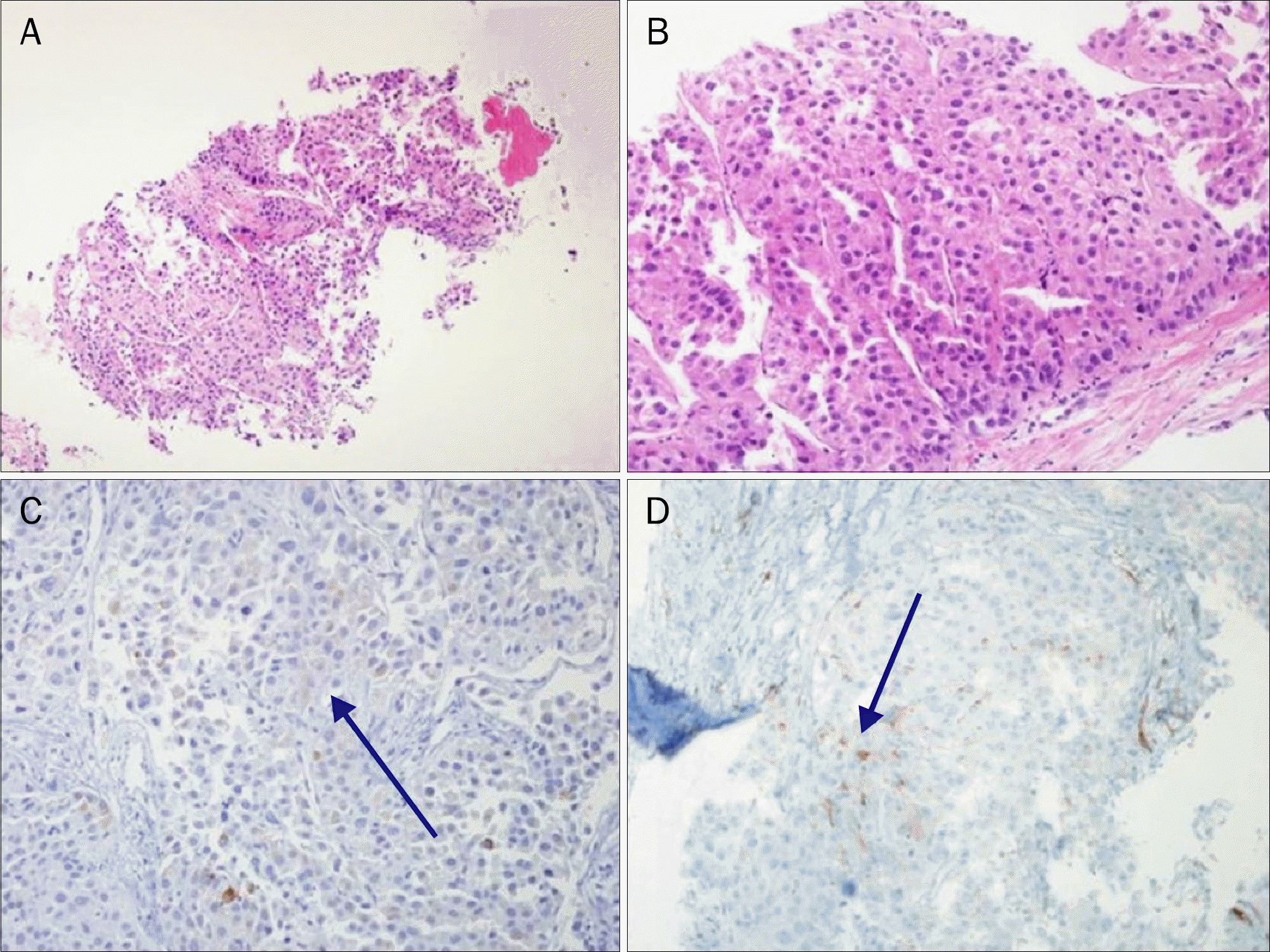
Fig. 5.
Abdomen CT scan after 2 months of sorafenib shows partial regression of the tumor mass (arrows).
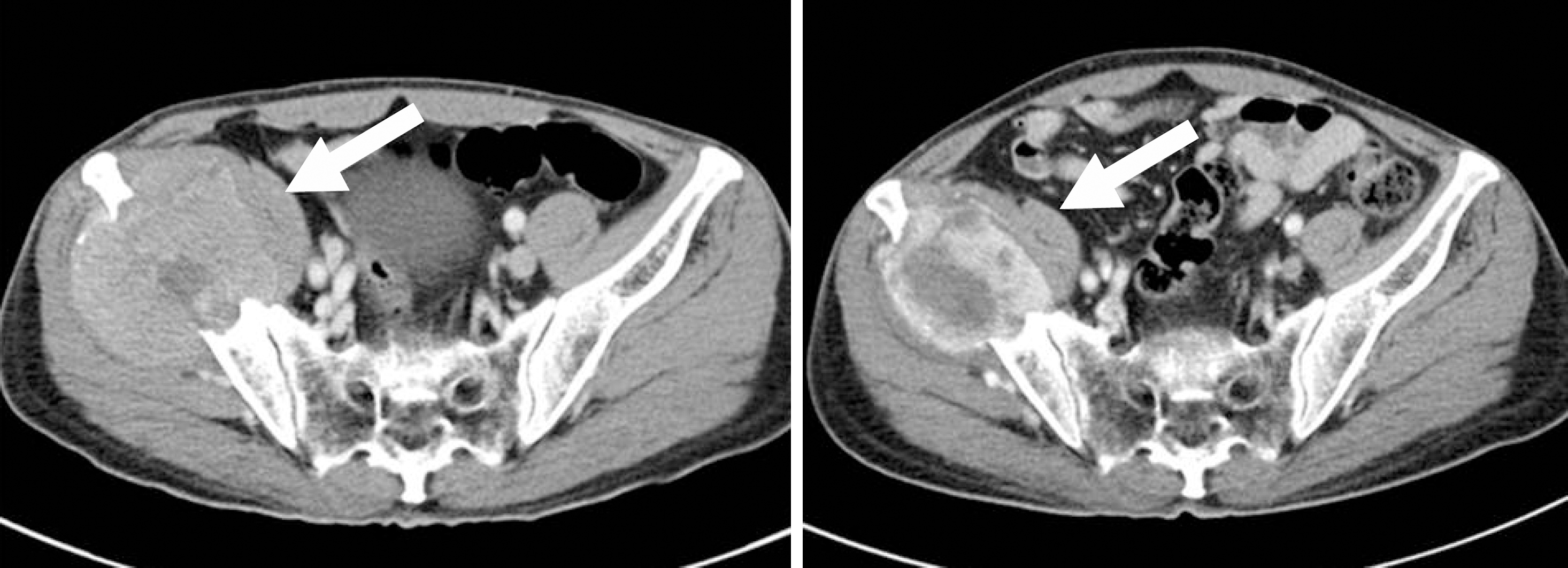
Table 1.
Motor Test of Upper Extremities
| Right | Left | |
|---|---|---|
| Shoulder | ||
| Flexor | 5 | 2 |
| Extensor | 5 | 4 |
| Elbow | ||
| Flexor | 5 | 3 |
| Extensor | 5 | 4 |


 PDF
PDF ePub
ePub Citation
 Print
Print


 XML Download