

 PDF
PDF ePub
ePub Citation
 Print
Print


Abstract
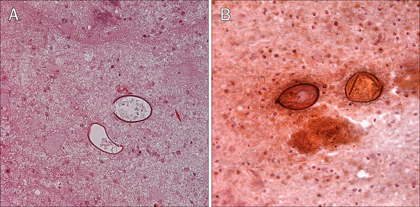
Paragonimiasis has been continuously decreasing in Korea. However, it still occurs by ingesting raw or incompletely cooked fresh water crab or crayfish. The diagnosis of paragonimiasis is challenging because of its rarity. It may be confused with other inflammatory disease or carcinomatosis. Endoscopic ultrasound-guided fine needle aspiration (EUS-FNA) has lower risk of complications such as bleeding, perforation than percutaneous fine needle aspiration. EUS-FNA is more accurate and popular method to find mucosal or submucosal tumors and the lesions of several organs. Benign and malignant tumors, infectious diseases have been diagnosed by EUS-FNA, but there was no report describing the use of EUS-FNA for diagnosing paragonimiasis. Herein, we present a 47-year-old male patient with paragonimiasis diagnosed by EUS-FNA. Imaging studies revealed mass lesions in the lung and peritoneal cavity, which was eventually confirmed as paragonimiasis using EUS-FNA.
Go to : 
References
1. Lee CH, Kim JH, Moon WS, Lee MR. Paragonimiasis in the abdominal cavity and subcutaneous tissue: report of 3 cases. Korean J Parasitol. 2012; 50:345–347.

2. Choi DW. Paragonimus and paragonimiasis in Korea. Kisaengchunghak Chapchi. 1990; 28(Suppl):79–102.
3. Catalano MF, Sial S, Chak A, et al. EUS-guided fine needle aspiration of idiopathic abdominal masses. Gastrointest Endosc. 2002; 55:854–858.
4. Costache MI, Iordache S, Karstensen JG, Săftoiu A, Vilmann P. Endoscopic ultrasound-guided fine needle aspiration: from the past to the future. Endosc Ultrasound. 2013; 2:77–85.
5. Jeon K, Koh WJ, Kim H, et al. Clinical features of recently diagnosed pulmonary paragonimiasis in Korea. Chest. 2005; 128:1423–1430.
6. Kim AY. Heterotopic paragonimiasis presented by intraabdominal masses. Korean J Gastroenterol. 2013; 61:351–353.
7. Kim TS, Han J, Shim SS, et al. Pleuropulmonary paragonimiasis: CT findings in 31 patients. AJR Am J Roentgenol. 2005; 185:616–621.
8. Kim KU, Lee K, Park HK, Jeong YJ, Yu HS, Lee MK. A pulmonary paragonimiasis case mimicking metastatic pulmonary tumor. Korean J Parasitol. 2011; 49:69–72.
9. Lee JJ, Choi CM, Kwon HH, et al. A case of pulmonary paragonimiasis mimicking lung cancer diagnosed by EBUS-TBNA. Korean J Med. 2013; 84:423–427.
10. Kim EY. Linear array endoscopic ultrasonography 1. Korean J Gastrointest Endosc. 2009; 38:1–8.
11. Eloubeidi MA, Tamhane A. Prospective assessment of diagnostic utility and complications of endoscopic ultrasound-guided fine needle aspiration. Results from a newly developed academic endoscopic ultrasound program. Dig Dis. 2008; 26:356–363.
12. Jenssen C, Dietrich CF. Endoscopic ultrasound-guided finenee-dle aspiration biopsy and trucut biopsy in gastroenterology – An overview. Best Pract Res Clin Gastroenterol. 2009; 23:743–759.
13. O'Toole D, Palazzo L, Arotçarena R, et al. Assessment of complications of EUS-guided fine-needle aspiration. Gastrointest Endosc. 2001; 53:470–474.
14. Carter JE, Nelson JJ, Eves M, Boudreaux C. Giardia lamblia infection diagnosed by endoscopic ultrasound-guided fine-nee-dle aspiration. Diagn Cytopathol. 2007; 35:363–365.
Go to :
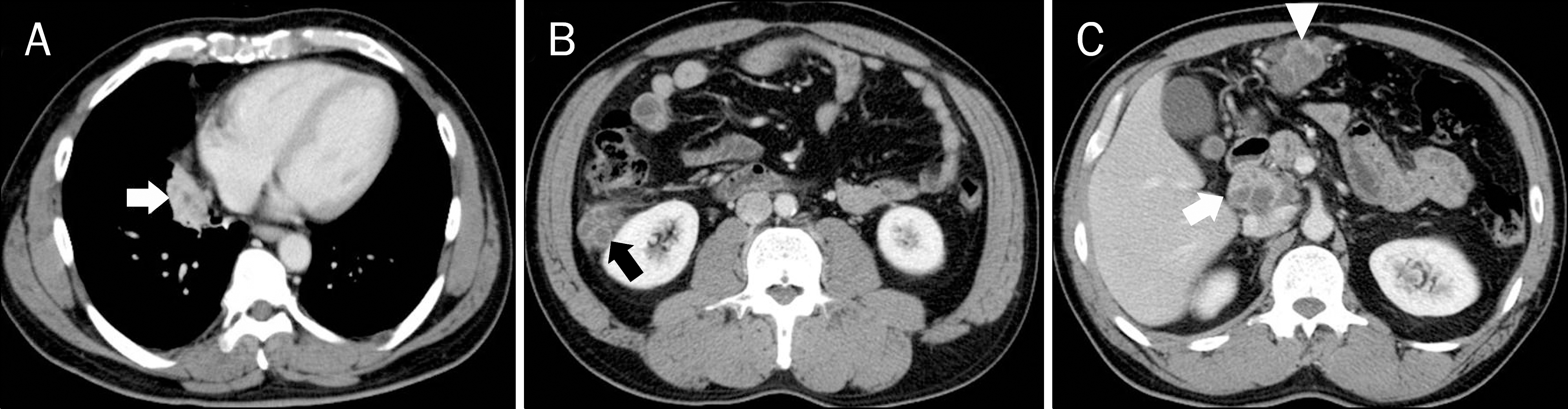 | Fig. 1.CT scans of chest and abdomen at admission. (A) At medial basal segment of right lower lobe, there is a 4.0 cm sized soft tissue density mass (arrow) harboring multifocal low attenuation areas. (B, C) On upper abdomen, there are three soft tissue density masses containing multiple necrotic low attenuation areas of bunch-of-grapes shape. A 3.5 cm sized mass (black arrow) is observed at right peri-renal space, a 4.5 cm sized mass (white arrowhead) at greater curvature side of stomach lower body along gastroepipoloic vessel, and a 4.6 cm sized mass (white arrow) at portocaval space. |
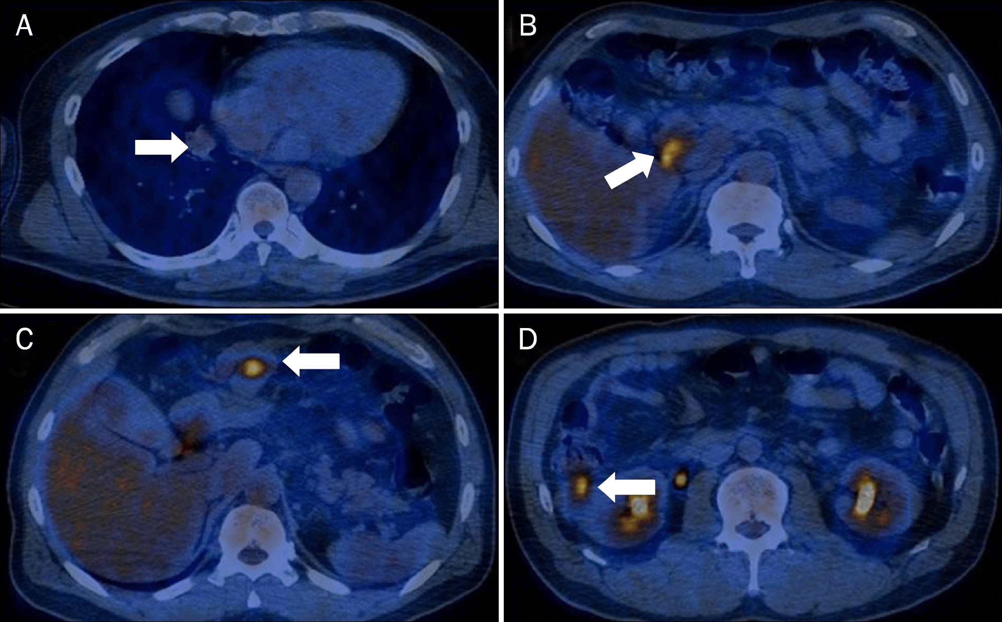 | Fig. 2.PET scan images. (A) PET scan reveals an ovoid nodular opacity (arrow) with mild F-18 fluorodeoxyglucose (FDG) uptake in the right lower lobe of lung (maximal standardized uptake value [SUV] 3.2). (B-D) PET scan reveals three low attenuation lesions (arrows) with increased FDG uptakes in the portocaval space, gastroepiploic area and right peri-renal space (maximal SUV 5.1). |
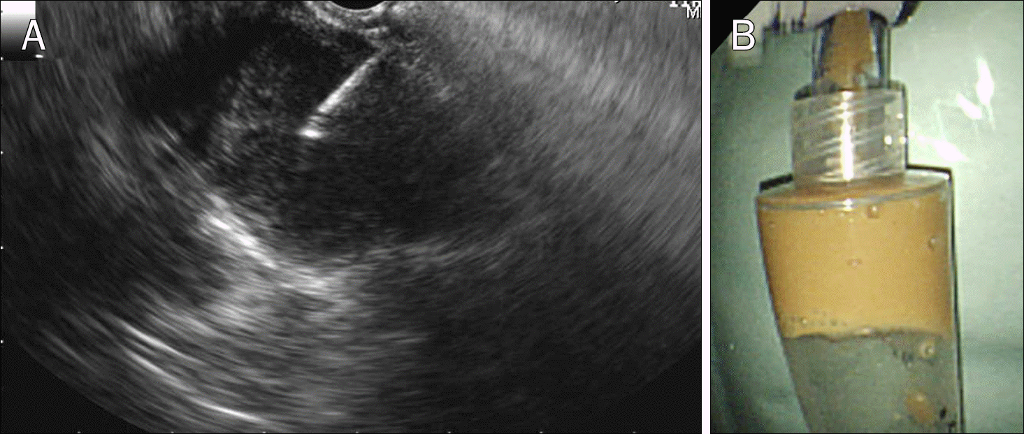 | Fig. 3.Endoscopic ultrasonography-guided fine needle aspiration findings. (A) On portocaval space, there was a heterogeneous hypoechoic 3 cm sized cystic mass with much debris. (B) After fine needle aspiration, muddy colored pus was obtained. |

 XML Download