

 PDF
PDF ePub
ePub Citation
 Print
Print


References
1. Jovanovic I, Knezevic S, Micev M, Krstic M. EUS mini probes in diagnosis of cystic dystrophy of duodenal wall in heterotopic pancreas: a case report. World J Gastroenterol. 2004; 10:2609–2612.

2. Surov A, Hainz M, Hinz L, et al. Case report: ectopic pancreas with pseudocyst and pseudoaneurysm formation. Clin Radiol. 2009; 64:734–737.
3. Schmitz H, Spelsberg F, Janson M. Heterotopic pancreatic cyst of the stomach wall. Leber Magen Darm. 1991; 21:33–35.
4. Eisenberger CF, Kropp A, Langwieler TE, Gocht A, Izbicki JR, Knoefel WT. Heterotopic pancreatitis: gastric outlet obstruction due to an intramural pseudocyst and hamartoma. Z Gastroenterol. 2002; 40:259–262.
5. Lee SL, Ku YM, Lee HH, Cho YS. Gastric ectopic pancreas complicated by formation of a pseudocyst. Clin Res Hepatol Gastroenterol. 2014; 38:389–391.
6. Mulholland KC, Wallace WD, Epanomeritakis E, Hall SR. Pseudocyst formation in gastric ectopic pancreas. JOP. 2004; 5:498–501.
7. Galloro G, Napolitano V, Magno L, et al. Diagnosis and therapeutic management of cystic dystrophy of the duodenal wall in heterotopic pancreas. A case report and revision of the literature. JOP. 2008; 9:725–732.
8. Park SS, Shin YM, Lim SW, et al. A case of symptomatic heterotopic pancreas with huge pseudocyst formation. Korean J Med. 2006; 70:706–710.
9. Wang JH, Lee JD, Kim CJ, et al. A case of gastric ectopic pancreas complicated by pancreatitis and pseudocyst formation. Korean J Gastrointest Endosc. 2003; 27:175–179.
10. Yen HH, Soon MS, Soon A. Heterotopic pancreas presenting as gastric submucosal cyst on endoscopic sonography. J Clin Ultrasound. 2006; 34:203–206.
11. Claudon M, Verain AL, Bigard MA, et al. Cyst formation in gastric heterotopic pancreas: report of two cases. Radiology. 1988; 169:659–660.
12. Fléjou JF, Potet F, Molas G, Bernades P, Amouyal P, Fékété F. Cystic dystrophy of the gastric and duodenal wall developing in heterotopic pancreas: an unrecognised entity. Gut. 1993; 34:343–347.
Fig. 1.
Initial abdomen CT (A, transverse view; B, coronal view). (A) The pancreas is slightly edematous without definite evidence of inflammations. (A, B) The stomach wall is thickened and 2 cm sized hypodense lesions are noted in the antrum (arrows).
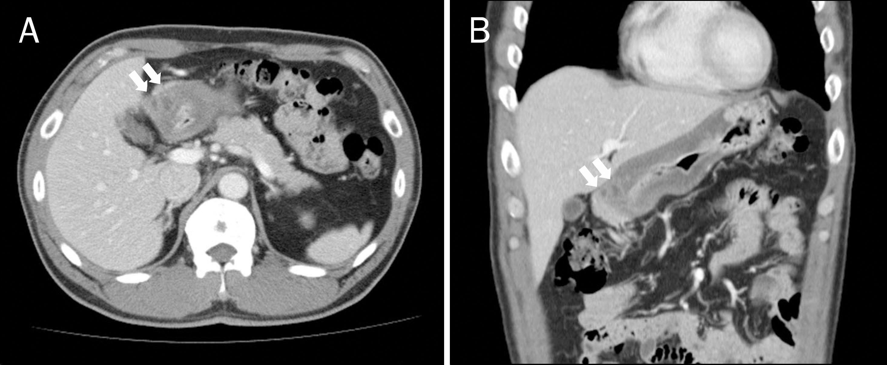
Fig. 2.
Endoscopic findings of upper gastrointestinal tract. (A) The wall of distal antrum is swollen with little erosive mucosal changes. (B) About 0.5 mm sized mucosal ulcer is noted in the pylorus.
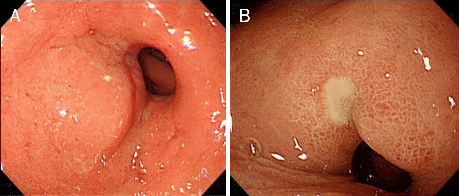
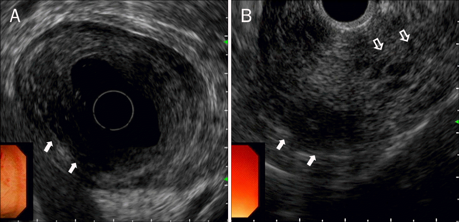
Fig. 3.
Endoscopic ultrasound findings.(A) On radial echoendoscopic view, entire wall of the stomach is swollen and little hypoechoic lesion (arrows) are noted. (B) On linear echoendo-scpic view, some inhomogenous-echo lesions (open arrows) with definite hypoechoic cystic lesions (closed arrows) are noted.
 XML Download